Indian Journal of Pharmacy and Pharmacology
Indian Journal of Pharmacy and Pharmacology (IJPP) is an open-access, peer-reviewed pharmacy journal, published quarterly, as print and online by the Innovative Education and Scientific Research Foundation (IESRF) since 2014. we will be publishing articles ‘Ahead of Print’ immediately upon acceptance of manuscript. In addition, the journal allows free access (Open Access) to its contents, which is likely to attract more readers and citations to articles published in journal.Manuscripts should be prepared in accordance with the author guidelines of the journal, which follow the recommendations...
Evaluation of anti-alzheimer activity of Lactuca sativa linn extract in scopolamine-induced amnesia in rat model

Abstract
Aim and Scope: The present study aimed to evaluate the anti-Alzheimer and memory-enhancing potential of the hydroalcoholic extract of Lactuca sativa Linn. leaves (H.E.L.S.) against scopolamine-induced amnesia in experimental rat models. The scope encompasses behavioral evaluation of spatial learning, memory retention, and histopathological assessment of neuronal protection.
Background: Alzheimer’s disease is a progressive neurodegenerative disorder characterized by severe memory impairment, cognitive decline, and structural brain changes such as neuronal loss and amyloid plaque deposition. Scopolamine, a muscarinic acetylcholine receptor antagonist, is widely used in preclinical research to induce temporary Alzheimer-like cognitive dysfunction and memory impairment in animal models. Material and
Methods: Animal Model: Adult Wistar albino rats divided into five treatment groups as Normal control, Disease control (Scopolamine-treated), Standard group (Donepezil 1mg/kg), Low dose treatment (H.E.L.S. 100mg/kg, p.o.), High dose treatment (H.E.L.S. 300mg/kg, p.o.). Induction: Alzheimer-like cognitive dysfunction was induced via intraperitoneal administration of scopolamine. Behavioral Assessments: Morris Water Maze (MWM)- To evaluate spatial learning and memory via escape latency. Elevated Plus Maze (EPM)- To measure learning and retention memory via open-arm exploration. Histopathological examination- Analysis of brain tissue sections to evaluate neuronal integrity and amyloid-related neurodegenerative changes.
Results: Behavioral Impairment: Scopolamine administration successfully induced cognitive dysfunction, marked by a significant increase in escape latency in MWM and decreased open-arm exploration in EPM. Cognitive Improvement: Oral administration of H.E.L.S. significantly reversed scopolamine-induced deficits in both behavioral models in a dose-dependent manner. Dose Comparison: The higher dose (300mg/kg) demonstrated a markedly superior neuroprotective effect compared to 100mg/kg, showing performance comparable to the standard drug, Donepezil. Histopathology: Brain tissue samples from H.E.L.S.-treated groups showed reduced neuronal degeneration and decreased amyloid plaque deposition relative to the disease control group.
Conclusion: The hydroalcoholic extract of Lactuca sativa Linn. leaves (H.E.L.S.) exhibits significant anti-Alzheimer activity by mitigating cognitive impairment and reducing structural brain damage (neuronal loss and amyloid plaques). These findings suggest that H.E.L.S., particularly at 300mg/kg, holds promise as a potential therapeutic candidate for managing neurodegenerative disorders like Alzheimer's disease.
1. Introduction
A neurodegenerative condition called Alzheimer's disease (AD) is typified by intracellular neurofibrillary tangles containing tau and extracellular plaques containing β amyloid (Aβ). A substantial amnestic cognitive impairment is the typical presentation of AD; non-amnestic cognitive impairment is a less prevalent manifestation. Though impairment in expressive speaking, visuospatial processing, and executive (mental agility) skills can occur, short-term memory difficulties are the most typical manifestation of AD. Many people with AD have a complicated genetic link, and the majority of instances of AD are not dominantly inherited. Patients with AD exhibit varying degrees of cognitive impairment. When objective cognitive testing results are not compromised, the initial signs may be a perceived deterioration in mental capacity.[1] A single cognitive domain or maybe many cognitive domains may be reduced to a low degree but functional abilities are mostly retained in mild cognitive impairment (MCI), which is the first symptomatic stage of cognitive impairment.[2] Dementia, on the other hand, is characterized by cognitive impairment severe enough to interfere with everyday functioning and impede independence. The hallmark clinical feature of AD is dementia with a progressive start and persistent progression accompanied by noticeable amnestic symptoms and indicators.[3]
|
Drug Used |
Class of Drug |
Mechanism of Action |
Side Effect |
|---|---|---|---|
|
Donepezil |
Cholinesterase inhibitors |
Donepezil inhibits acetylcholinesterase, leading to increased acetylcholine levels in the synaptic cleft. |
Nausea, diarrhea, insomnia, muscle cramps, fatigue, and anorexia. |
|
Rivastigmine |
Cholinesterase inhibitors |
Inhibits both acetylcholinesterase and butyrylcholinesterase, leading to increased acetylcholine levels. |
Nausea, vomiting, loss of appetite, and weight loss. |
|
Galantamine |
Cholinesterase inhibitors |
Galantamine is a reversible cholinesterase inhibitor and also modulates nicotinic receptors to enhance cholinergic function. |
Nausea, vomiting, diarrhea, dizziness, and weight loss. |
|
Tacrine |
Cholinesterase Inhibitors |
Tacrine inhibits acetylcholinesterase, the enzyme responsible for breaking down acetylcholine in the synaptic cleft. |
Hepatotoxicity nausea, vomiting, diarrhea, Bradycardia |
|
Memantine |
NMDA Receptor Antagonist |
Memantine blocks NMDA receptors, reducing abnormal glutamate activity that can lead to neuronal damage. |
Dizziness, headache, constipation, confusion, and hypertension. |
When AD was first identified, it was thought to be a clinicopathological entity, which meant that if a patient had amnestic dementia and all other possible causes had been ruled out, AD pathology was likely to be the reason.[4] However, the definition of AD has changed to reflect a neurobiological illness that affects several parts of cognition due to improved clinical complexity and biomarkers of AD, such as cerebrospinal fluid (CSF) and PET indicators for tau and Aβ. Notably, there is a growing understanding of the connection between AD as well as additional causes of cognitive decline. AD is a synaptic dysfunction condition with a preference for a cognitively expressive brain that includes failures of the macro-, molecular, and cellular cortical circuitry systems. When it comes to connecting findings concerning neuropathology, genetics, cell biology, and the clinical presentations of AD, synaptic pathophysiology is an appealing issue. Positive or "overt" lesions that are visible under a microscope, such as tau-containing neurofibrillary tangles, Aβ-containing plaques, activated glia, or larger endosomes, can be used to describe the pathophysiology of AD. Alternatively, AD may be seen as a representation of negative (or "covert") processes, such as the loss of neurons, synaptic homeostasis, or the integrity of neural networks. Although the biology of tau protein and amyloid precursor protein is extensively discussed in this primer, the amyloid cascade hypothesis,[5] which places these proteins at the forefront, ignores the numerous other hidden mechanisms that have been proposed as being important but cannot all be discussed here.[6][7] Although a great deal of research has been done, there is still much to understand about how the processes underlying AD generate synaptic and neuronal loss, which are the most likely sources of cognitive impairment.
Currently, medications used to treat Alzheimer's disease (AD) mostly target the illness's symptoms rather than its cause or progression. Cholinesterase inhibitors and NMDA receptor antagonists are the two primary medication classes that are used. The commonly used medications, their modes of action, and related side effects are listed in Table 1.
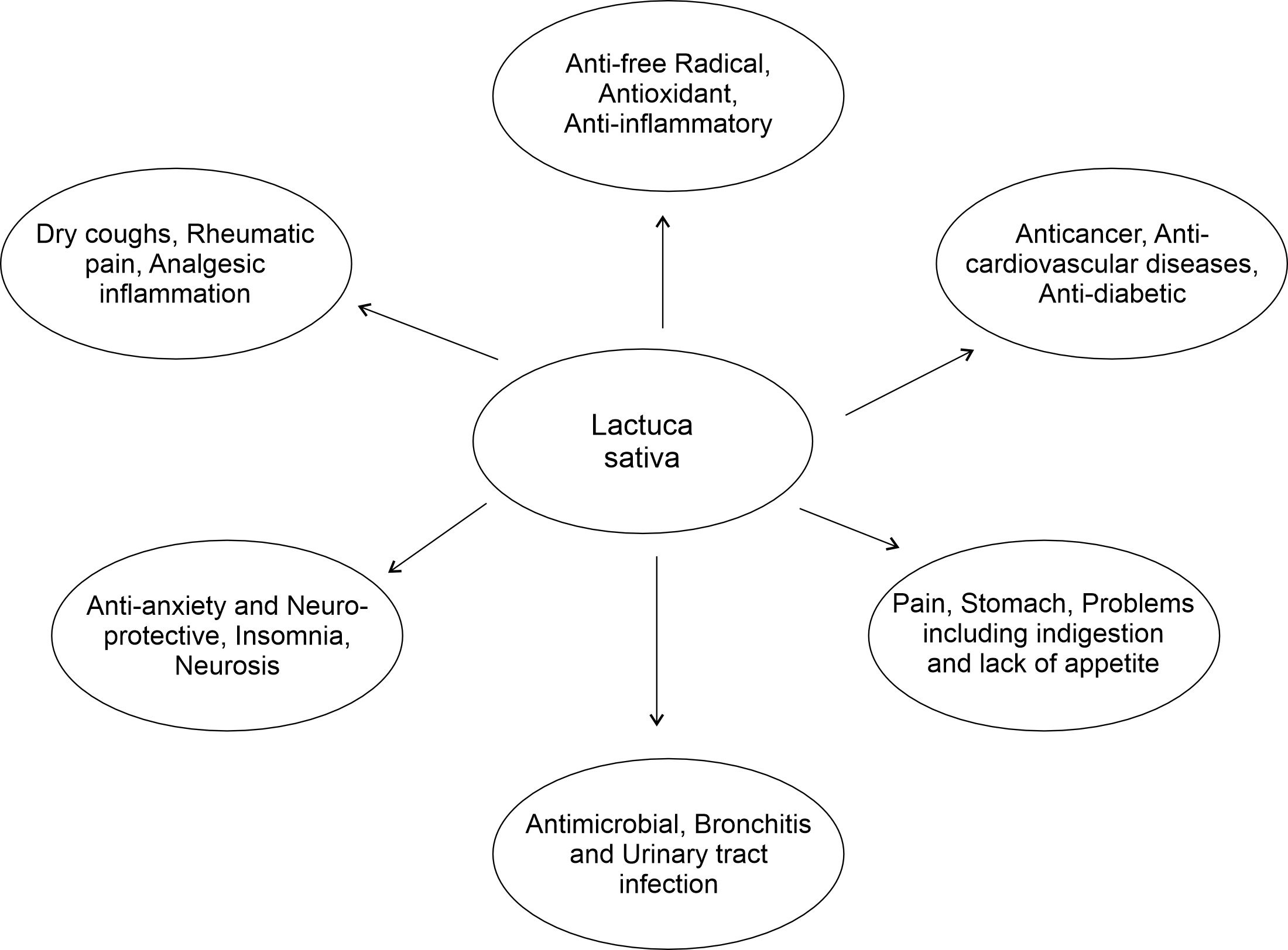
Lettuce is considered the most consumed salad. Vegetables rich in phytonutrients. The organic compounds and antioxidant activity of lettuce depend on the type but may also vary among similar breeds, breeding conditions, and genotypes. It also affects the structure of biologically active compounds in plants. Lettuce (Lactuca sativa L.) belongs to the Asteraceae family and originated in the Mediterranean region. It is a successful and abundant plant, which is found all over the world. Front Cultivated lettuce appears as a plant in texts as early as 2680 BC. Asteraceae is considered to be the most diverse plant family approximately 23,000–30,000 number of species. It is rich in calcium, iron, vitamin A.[8][9] The whole plant is rich in a milky sap, sap contains lactucarium which is used in medicine for sedative, digestive, diuretic, narcotic properties. It is taken internally in the treatment of insomnia, anxiety, neurosis, dry coughs, rheumatic pain, etc.[10] The whole plant has been used as a traditional medicine for the treatment of stomach problems, stimulate digestion and to enhance appetite and relieve inflammation ([Figure 1]).[11] This study revealed that lettuce extract has the potential for memory enhancement against scopolamine-induced memory loss.
2. Materials and Methods
2.1. Chemicals and reagents
All the chemicals and reagents used were of analytical reagent grade and were obtained from Labware Chemicals, Latur, Maharashtra, India.
2.2. Plant collection and authentication
The Lactuca sativa was collected from local Latur District, Maharashtra State of India. The plant was authenticated by Dr. A. Benniamin, Scientist In-charge, Botanical Survey of India, Western Regional Centre Pune (M.H.) with wide reference number - BSI/WRC/Tech./2024/ JVD-43.
2.3. Plant extraction
Leaf of Lactuca sativa Linn was rinsed well with tap water and distilled water and kept under shade for drying for 7 days. Dried material coarsely powdered using mortar and pestle and further reduced to powder using an electric blender and stored in air tight glass container. The powder was subjected to solvent extraction so that 30 g of powder was extracted in 300mL of Ethanol:Water (70:30) for 5 hrs. After drying the extract was stored in the container.[12]
2.4. Qualitative analysis
2.4.1. Physical characteristics
The physical characteristics of Lactuca sativa linn leaf extract involves physical appearance and solubility.
2.4.2. FT-IR analysis
FTIR spectroscopy was used to investigate the drug for its functional groups at range 4000cm-1 to 400cm-1. The FTIR study performed using Perkin Elmer FTIR.
2.4.3. Phytochemical screening
The crude extract was evaluated for the presence of various phytoconstituent such as carbohydrates, proteins, alkaloids, glycosides, terpenoids, steroids, flavonoids, tannins and saponins.[13][14]
2.5. Experimental study
2.5.1. Selection and procurement of animals
Prior to starting the experimentation, approval from the Institutional Animal Ethics Committee (IAEC) was acquired for the study plan. A total of 30 Wistar rats, weighing between 180 and 200g, were procured from the Crystal biological solution, Pune, Maharashtra, India, for carrying out experimental investigation.
2.5.2. Dosage preparations
Lactuca sativa linn leaf extract: Lactuca sativa Linn Leaf extract at doses 100mg/kg and 300mg/kg were selected for the study. Lactuca sativa Linn Leaf extract was suspended in 1% tween 80 solution and administered by oral route. Donepezil: It is administered to SCOP + Donepezil group in the dose of 1mg/kg p.o. in normal saline (2ml/100gm, p.o.). Donepezil is freely soluble in water and partially soluble in organic solvents so it was administered in distilled water. Scopolamine: Hyoscine butyl bromide (20mg/ml) diluted in Bacteriostatic water for injection was given to all groups except control group.
2.5.3. Experimental Procedure
After weeks of habituation period, rats were randomly assigned to 5 groups. Each group consisted of 6 rats. All treatments were given every day at the same time of the day. Behavioral procedures were performed between 9 a.m. and 4 p.m. Control group (Group 1): Received normal saline (p.o.) for 15 days from day 1–6 and after that from day 9–17. SCOP group (Group 2): Received normal saline (p.o.) for 15 days from day 1–6 and after that from day 9–17. Received 1mg/kg scopolamine (i.p) to rats on day 9–17. SCOP + Donepezil group (Group 3): Received 1mg/kg of Donepezil (p.o.) daily for 15 days from day 1–6 and day 9–17. From day 9, 1mg/kg scopolamine (i.p) was administered after half an hour of donepezil administration. SCOP + Test1group (Group 4): Received 100mg/kg/day (p.o.) for daily for 15 days from day 1–6 and day 9–17. From day 9–17, 1mg/kg scopolamine (i.p) was administered after half an hour of test 1 administration. SCOP + Test 2 group (Group 5): Received 300 mg/kg/day for orally once daily for 15 days from day 1–6 and day 9–17. From day 9–17, scopolamine was administered after half an hour of test 2 administration.
All experiments were performed in a balanced design (six animals per group) to avoid being influenced by order and time. The behavioral studies were divided into two phases, namely Phase I and Phase II. During Phase I, all experimental groups received their respective oral pretreatments for six consecutive days before being subjected to a battery of behavioral tests. Behavioral assessments were performed from Day 6 to Day 8 using the Morris water maze and elevated plus maze tests. Scopolamine (1mg/kg B.W., i.p.) was administered 30 minutes before each behavioral test to induce amnesia. During Phase II study, amnesia was induced in all the groups except the control group by daily intraperitoneal injections of scopolamine (1mg/kg) for 9 days after extract pre-treatment (Day 9–17). Thirty minutes prior to the administration of scopolamine, Morrise water test was conducted on Day 10 and Day 15, and EPM was carried out on Days 11 and 12, and Days 16 and 17. At the end of the experiment, the rats were sacrificed, and their brains were isolated for histopathology.
2.5.3.1. Behavioral assessment
Morris water maze (MWM) test: The MWM task was used to gauge rat’s capacity for spatial long-term memory. The MWM consisted of a black circular pool (1.2m diameter, 50cm height). The pool was in a quiet room with several environmental cues (colorful shapes). Throughout the testing days, the pool and the environmental cues were kept the same. Water was added to the pool at a temperature of 22 ± 2°C. The MWM was almost evenly split into four quadrants: I, II, III, IV. A platform of black color (9cm in diameter and 20 cm height) was centered in 4th quadrant 1.5cm below the surface of water. The water was kept clear. The black color of pool and platform made it hard for rats to recognize the platform easily. The platform position was kept unaltered throughout the experimentation. Three phases: pre-acquisition, Acquisition and Retention were carried out. In each phase four trials per day were taken for each rat. After each trial rats were dried with a towel. The Morris water maze test was conducted on Days 6, 10, and 15. Scopolamine (1mg/kg B.W., i.p.) was administered 30 minutes before each trial to induce amnesia. All trials were recorded using a digital video camera. The platform was taken out of the water during the retention phase. The latency time (time required to arrive at the formal platform’s location and the amount of time spent in the target quadrant, over the course of 60s, was noted.[15] Elevated plus maze (EPM)test: Rodent learning and memory can be assessed during the Elevated Plus Maze (EPM), an exteroceptive behavioral model. Two open arms (50 × 10cm) and closed arms (50 × 10 × 40cm) that extended from a single central platform (10 × 10cm) made up the EPM, which was made of medium density fiber board with a matte black acrylic surface. The maze was elevated to a height of 50cm above the floor level. Each rat was positioned at the end of an open arm, facing away from the central platform in the habituation phase and were allowed to explore the maze for 5 min. Rats were positioned on the central platform in the acquisition phase, on first day of the test, 30 minutes to the drug treatments. Transfer latency (TL) was defined as the amount of time it took an animal with all four limbs to enter any one of the closed arms. The animal that did not enter into one of the closed arms were gently pushed into one at the end of the 60 second trial, at which point the TL was determined to be 60 seconds. For a further two minutes, the rat was free to explore the maze. Expect for the control group, retention latency was measured after 24 hrs. The scopolamine administration, during the retention phase. Memory progress can be shown by a decrease in TL values on the second day of the test compared to the first day.[16]
2.6. Histopathological studies
On day 18, rats were sacrificed and brains were excised immediately. Brain tissues were washed with ice-cold normal saline and fixed in 10% formalin for 24–48 h. The hippocampal and cortical regions were processed by paraffin embedding technique, sectioned at 5 µm thickness using a rotary microtome, and stained with Hematoxylin and Eosin (H&E). The stained sections were examined under a light microscope for histopathological changes such as neuronal degeneration, pyknosis, amyloid aggregation, and degeneration of pyramidal cells. Photomicrographs were captured for comparative evaluation of neuroprotective activity.
2.7. Statistical analysis
All data were expressed as mean ± SEM (n = 6). Statistical analysis was performed using one-way analysis of variance (ANOVA) followed by Tukey's multiple comparison test using Graph-pad prism software (Vesion 9.0). p value of < 0.05 was considered statistically significant.
3. Results
3.1. Physical characteristics (Table 2)
|
Physical characteristics |
|
|---|---|
|
Color |
Dark Green |
|
Odor |
Earthy smell |
|
Texture |
Semi solid |
|
**Solubility** |
|
|
Water |
Slightly soluble |
|
Ethanol |
Soluble |
|
Methanol |
Soluble |
|
Tween 80 |
Soluble |
3.2. FTIR analysis
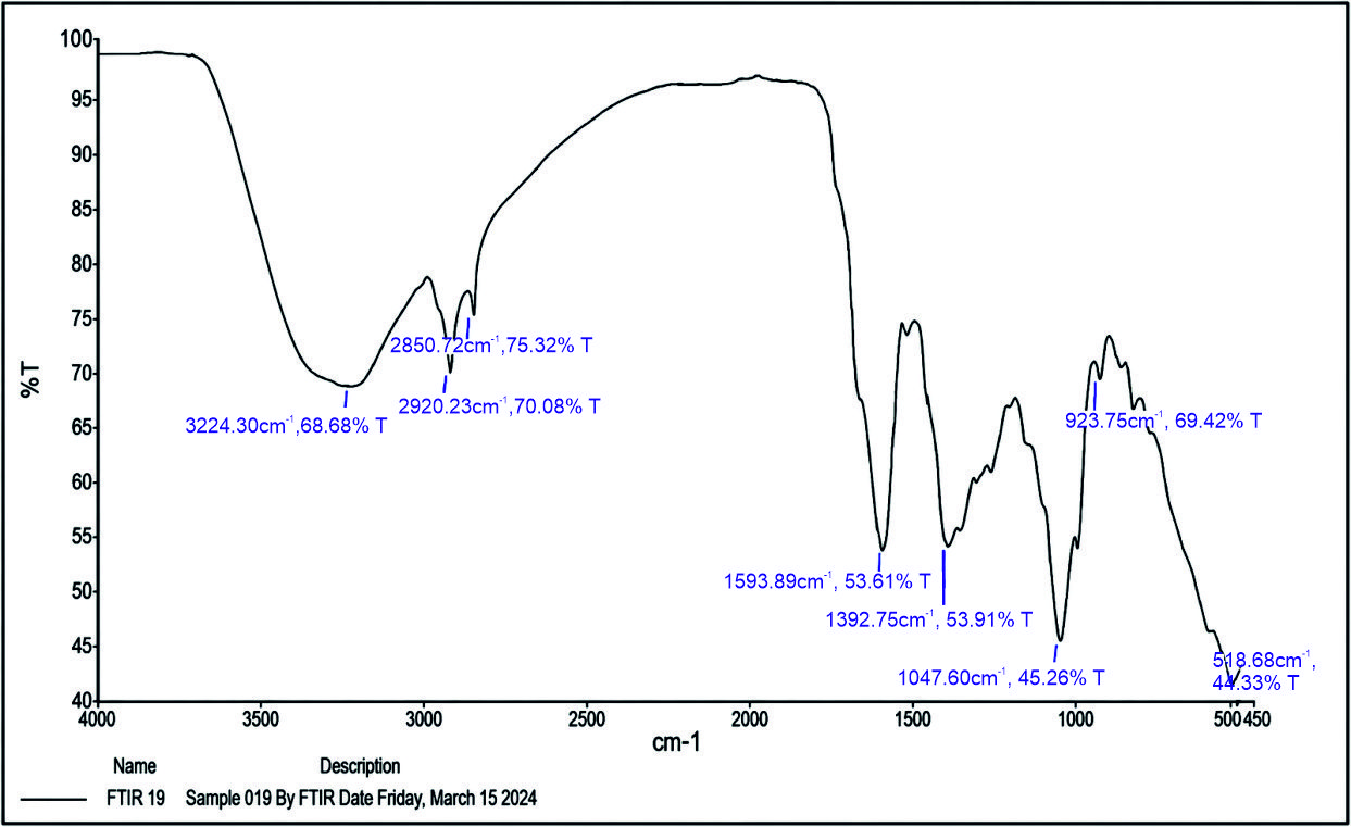
The FTIR analysis of H.E.L.S. revealed the presence of various functional groups corresponding to different phytochemical constituents. A broad absorption peak observed at 3224.30cm⁻¹ indicated the presence of hydroxyl (–OH) groups, suggesting phenolic compounds. The peak at 2920.23cm⁻¹ corresponded to aromatic C–H stretching, while the band at 2850.72cm⁻¹ represented aliphatic C–H stretching vibrations. A strong absorption at 1593.89cm⁻¹ indicated aromatic C=C stretching, confirming the presence of aromatic compounds such as flavonoids. The peak at 1392.75cm⁻¹ was attributed to aliphatic C–C stretching, whereas the absorption at 1047.60cm⁻¹ corresponded to ether functional groups, indicating the presence of glycosidic or polysaccharide structures. Overall, the FTIR spectrum confirmed the presence of key bioactive phytoconstituents such as phenols, flavonoids, alkaloids, and glycosidic compounds in H.E.L.S., which may contribute to its observed neuroprotective and anti-Alzheimer activity ([Figure 2]).
3.3. Phytochemical screening
The screening the phytochemical present in Lactuca sativa linn leaf extract shown in Table 3.
3.4. Experimental study
3.4.1. Effect of Lactuca sativa linn leaf extract on working and spatial reference memory in the Morris Water Maze Test
The results showed that the disease control group treated with normal saline and scopolamine exhibited a significant increase in escape latency time on Day 6 (23.6 ± 1.32 sec), Day 10 (32.1 ± 2.14 sec), and Day 15 (33.5 ± 1.12 sec) compared to the control group, indicating impaired memory and learning. Treatment with Donepezil (1mg/kg, p.o.) significantly reduced the escape latency time to 19.4 ± 3.35 sec on Day 6, 18.6 ± 4.11 sec on Day 10, and 18.3 ± 2.13 sec on Day 15, demonstrating improved cognitive performance. Similarly, H.E.L.S. at 100mg/kg showed a reduction in escape latency time with values of 22.6 ± 4.21 sec, 21.8 ± 2.17 sec, and 21.2 ± 1.16 sec on Days 6, 10, and 15 respectively. The higher dose of H.E.L.S. (300mg/kg) produced a more pronounced effect, reducing escape latency time to 19.8 ± 2.26 sec on Day 6, 18.9 ± 4.21 sec on Day 10, and 18.6 ± 3.19 sec on Day 15, comparable to the standard drug Donepezil ([Table 4]).
|
Phytoconstituents |
Result |
|---|---|
|
Alkaloids |
+ |
|
Carbohydrates |
+ |
|
Polyphenols |
+ |
|
Flavonoids |
+ |
|
Terpenoids |
+ |
|
Sterols |
+ |
|
Tannin |
+ |
|
Glycosides |
- |
|
Cardiac Glycoside |
- |
|
*+ indicates Presence, - indicates Absence |
|
Group |
Treatment |
Escape Latency Time (sec) ± SEM |
||
|---|---|---|---|---|
|
**Day 6** |
**Day 10** |
**Day 15** |
||
|
Control |
Normal saline (p.o.) |
23.5 ± 2.31 |
22.4 ± 3 .26 |
20.9 ± 2 .21 |
|
Disease control |
Normal saline (p.o.) + scopolamine (i.p.) |
23.6 ± 1.32### |
32.1 ± 2.14### |
33.5 ± 1.12### |
|
Standard |
Donepezil 1mg/kg (p.o.) |
19.4 ± 3.35*** |
18.6 ± 4.11*** |
18.3 ± 2.13*** |
|
H.E.L.S. 100mg/kg |
H.E.L.S. 100mg/kg (p.o.) |
22.6 ± 4.21** |
21.8 ± 2.17*** |
21.2 ± 1.16*** |
|
H.E.L.S. 300mg/kg |
H.E.L.S. 300mg/kg (p.o.) |
19.8 ± 2.26*** |
18.9 ± 4.21*** |
18.6 ± 3.19*** |
|
Day |
Treatment |
Transfer latency (Sec) ± SEM |
|---|---|---|
|
7 |
Normal saline (p.o.) |
22.56 ± 1.36 |
|
Normal saline (p.o.) + scopolamine (i.p.) |
28.54 ± 1.89### |
|
|
Donepezil 1mg/kg (p.o.) |
25.34 ± 1.58*** |
|
|
H.E.L.S. 100mg/kg (p.o.) |
28.34 ± 2.09*** |
|
|
H.E.L.S. 300mg/kg (p.o.) |
26.65 ± 1.36*** |
|
|
8 |
Normal saline (p.o.) |
20.67 ± 1.54 |
|
Normal saline (p.o.) + scopolamine (i.p.) |
32.45 ± 2.11### |
|
|
Donepezil 1mg/kg (p.o.) |
18.65 ± 1.05*** |
|
|
H.E.L.S. 100mg/kg (p.o.) |
22.12 ± 1.78*** |
|
|
H.E.L.S. 300mg/kg (p.o.) |
20.34 ± 1.65*** |
|
Day |
Treatment |
Transfer latency (Sec) ± SEM |
|---|---|---|
|
11 |
Normal saline (p.o.) |
18.45 ± 0.99 |
|
Normal saline (p.o.) + scopolamine (i.p.) |
35.65 ± 2.45### |
|
|
Donepezil 1mg/kg (p.o.) |
16.56 ± 2.06*** |
|
|
H.E.L.S. 100mg/kg (p.o.) |
20.18 ± 1.78*** |
|
|
H.E.L.S. 300mg/kg (p.o.) |
18.87 ± 1.55*** |
|
|
12 |
Normal saline (p.o.) |
16.78 ± 2.33 |
|
Normal saline (p.o.) + scopolamine (i.p.) |
38.78 ± 2.11### |
|
|
Donepezil 1mg/kg (p.o.) |
14.77 ± 2.75*** |
|
|
H.E.L.S. 100mg/kg (p.o.) |
18.22 ± 1.15*** |
|
|
H.E.L.S. 300mg/kg (p.o.) |
15.99 ± 2.25*** |
|
Day |
Treatment |
Transfer latency (Sec) ± SEM |
|---|---|---|
|
16 |
Normal saline (p.o.) |
15.55 ± 1.75 |
|
Normal saline (p.o.) + scopolamine (i.p.) |
40.42 ± 1.85### |
|
|
Donepezil 1mg/kg (p.o.) |
12.17 ± 1.86*** |
|
|
H.E.L.S. 100mg/kg (p.o.) |
15.22 ± 2.36*** |
|
|
H.E.L.S. 300mg/kg (p.o.) |
14.72 ± 1.65*** |
|
|
17 |
Normal saline (p.o.) |
12.84 ± 2.08 |
|
Normal saline (p.o.) + scopolamine (i.p.) |
45.55 ± 1.91### |
|
|
Donepezil 1mg/kg (p.o.) |
9.52 ± 1.95*** |
|
|
H.E.L.S. 100mg/kg (p.o.) |
14.23 ± 2.65*** |
|
|
H.E.L.S. 300mg/kg (p.o.) |
10.22 ± 1.78*** |
3.4.2. Effect of Lactuca sativa linn leaf extract on working and spatial reference memory in the Elevated plus maze (EPM) test
The Elevated Plus Maze (EPM) results on Days 7 and 8 revealed that the scopolamine-treated disease control group exhibited a significant increase in transfer latency compared with the normal control group, indicating impaired learning and memory. Treatment with Donepezil (1mg/kg, p.o.) significantly reduced the transfer latency. H.E.L.S. at 100mg/kg also decreased the transfer latency, whereas H.E.L.S. at 300mg/kg produced a more pronounced reduction, with effects comparable to those of Donepezil ([Table 5]).
On Days 11 and 12, the scopolamine-treated disease control group exhibited a significant increase in transfer latency compared with the normal control group, indicating impaired learning and memory. Treatment with Donepezil (1mg/kg, p.o.) markedly reduced the transfer latency. H.E.L.S. at 100mg/kg produced moderate improvement, whereas the 300mg/kg dose resulted in a greater reduction in transfer latency, indicating enhanced cognitive performance comparable to that of Donepezil ([Table 6]).
On Days 16 and 17, the scopolamine-treated disease control group exhibited the highest transfer latency among all experimental groups, indicating severe cognitive impairment. Administration of Donepezil (1mg/kg, p.o.) significantly shortened the transfer latency. H.E.L.S. treatment produced a dose-dependent improvement, with the 300mg/kg dose producing a greater reduction in transfer latency than the 100mg/kg dose and exhibiting cognitive-enhancing effects comparable to those of the standard drug, Donepezil ([Table 7]).
3.5. Histopathological analysis
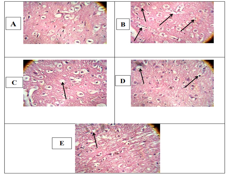
The normal control group showed normal architecture of the brain cortex without any evidence of neurodegeneration or necrosis. The disease control group treated with scopolamine exhibited severe neuronal degeneration, pyknosis in hippocampal sections, aggregation of amyloid plaques, and focal degeneration of pyramidal cells. The standard drug donepezil-treated group showed marked protection with only minimal degeneration and pyknosis along with mild amyloid aggregation. The H.E.L.S. treated groups showed dose-dependent neuroprotective effects, where the low dose (100mg/kg) demonstrated moderate improvement, while the high dose (300mg/kg) exhibited mild degeneration and minimal amyloid aggregation, indicating significant protection against scopolamine-induced neurodegenerative changes ([Figure 3]).
4. Discussion
Alzheimer’s disease (AD) is a progressive neurodegenerative disorder characterized by deterioration of memory, cognition, thinking ability, and behavioral functions, ultimately interfering with daily activities. The development of memory impairment and dementia has become a major global health concern, particularly among the aging population.[17] Several medicinal plants have been reported to possess neuroprotective and memory-enhancing activities through multiple mechanisms such as acetylcholinesterase (AChE) inhibition, antioxidant action, modulation of neurotrophic factors, and prevention of neuronal cell death. Therefore, natural product-based therapies, nutraceuticals, and plant-derived compounds have gained increasing attention for the management of age-related neurodegenerative disorders including AD.[18]
In the present study, scopolamine-induced amnesia was employed as an experimental model to evaluate the anti-Alzheimer potential of hydroalcoholic extract of Lactuca sativa Linn leaves using the Morris Water Maze and Elevated Plus Maze models. Scopolamine is known to impair cholinergic neurotransmission by blocking muscarinic receptors, thereby producing learning and memory deficits similar to those observed in Alzheimer’s disease. The results of the present investigation demonstrated that HELS significantly improved memory and cognitive functions in scopolamine-treated animals. Significant improvement was observed in escape latency during the MWM test and in transfer latency during the EPM test. The standard drug, Donepezil produced the most prominent improvement in all behavioral parameters, whereas H.E.L.S. treatment significantly improved cognitive performance, with the 300mg/kg dose exhibiting greater anti-amnesic activity than the 100mg/kg dose.
Among the tested doses, H.E.L.S. 300 mg/kg exhibited superior efficacy and produced results comparable to Donepezil, suggesting dose-dependent neuroprotective activity. The treated groups showed improved learning acquisition, memory retention, and retrieval compared with the disease control group. Furthermore, histopathological studies revealed that H.E.L.S. treated groups exhibited improved neuronal architecture and regenerative changes compared with the scopolamine-treated animals, indicating its protective effect against neuronal degeneration.
The neuroprotective and anti-Alzheimer activity of H.E.L.S. may be attributed to the presence of various bioactive phytoconstituents identified during phytochemical screening, including flavonoids, alkaloids, carotenoids, terpenoids, sterols, and tannins. Flavonoids are well known for their potent antioxidant and free radical scavenging properties, which help reduce oxidative stress and neuronal damage associated with AD.[19] Steroidal compounds and terpenoids may contribute to neuroprotection through anti-inflammatory and membrane-stabilizing actions, while alkaloids may enhance cholinergic neurotransmission by inhibiting acetylcholinesterase activity. The antioxidant phytoconstituents present in HELS may further protect neuronal cells from oxidative damage and apoptosis, thereby improving cognitive performance and memory function.[20]
The present study findings suggest that H.E.L.S. possesses significant anti-Alzheimer and neuroprotective potential, possibly mediated through antioxidant activity, cholinergic enhancement, and prevention of neuronal degeneration. The higher dose of H.E.L.S. 300 mg/kg revealed better efficacy and may serve as a promising natural therapeutic candidate for the management of Alzheimer’s disease.
5. Conclusion
The present study demonstrated that hydroalcoholic extract of Lactuca sativa linn. (H.E.L.S.) significantly improved scopolamine-induced memory impairment and behavioral deficits in experimental animals. H.E.L.S. exhibited notable neuroprotective and anti-Alzheimer activity, particularly at 300 mg/kg, comparable to Donepezil. The activity may be attributed to flavonoids, alkaloids, terpenoids, and antioxidant phytoconstituents present in the extract. Further studies may support its development into an effective herbal formulation for Alzheimer’s disease management.
6. Authors Contribution
Mahesh Bandappa Manke: Conceptualization, Data curation, Formal analysis, Investigation, Methodology, Project administration, Software, Writing – original draft. Vinayaksing Sumersing Suryavanshi: Conceptualization, Data curation, Investigation, Methodology, Writing – original draft. Rachana Vijayanand Hallale: Formal analysis, Investigation. Padmaja Sidram Giram: Data curation, Formal analysis, Supervision, Writing – review editing. Omprakash Gadgeappa Bhusnure: Supervision, Visualization, Writing – review editing.
7. Source of Funding
None.
8. Conflict of Interest
None.
9. Ethical Approval
The study was approved by the Institutional Animal Ethics Committee (IAEC) of Channabasweshwar Pharmacy College, Latur, Maharashtra. (Ref. No. CPCSEA/CBPL/AH/86).
References
- Jessen F, Amariglio RE vBM, Breteler M, Ceccaldi M CG. A conceptual framework for research on subjective cognitive decline in preclinical Alzheimer's disease. Alzheimers Dement. 2014;10(6):844-52. [Google Scholar] [Crossref]
- Petersen R. Mild cognitive impairment as a diagnostic entity. J Intern Med. 2004;256(3):183-94. [Google Scholar] [Crossref]
- McKhann G, Knopman D, Chertkow H, Hyman B, Jack Jr C, Kawas C. The diagnosis of dementia due to Alzheimer's disease: recommendations from the National Institute on Aging-Alzheimer's Association workgroups on diagnostic guidelines for Alzheimer's disease. Alzheimers Dement. 2011;7(3):263-9. [Google Scholar] [Crossref]
- Petersen R, Lopez O, Armstrong M, Getchius T, Ganguli M, Gloss D. Practice guideline update summary: Mild cognitive impairment [RETIRED]: Report of the Guideline Development, Dissemination, and Implementation Subcommittee of the American Academy of. Neurology. 2018;90(3):126-135. [Google Scholar] [Crossref]
- Golde T, DeKosky S, Galasko D. Alzheimer's disease: The right drug, the right time. Science. 2018;362(6420):1250-1. [Google Scholar] [Crossref]
- Herrup K. The case for rejecting the amyloid cascade hypothesis. Nat Neurosci. 2015;18(6):794-9. [Google Scholar] [Crossref]
- Zlokovic B, Gottesman R, Bernstein K, Seshadri S, McKee A, Snyder H. Vascular contributions to cognitive impairment and dementia (VCID): A report from the 2018 National Heart, Lung, and Blood Institute and National Institute of Neurological Disorders and Stroke Workshop. Alzheimers Dement. 2018;16(12):1714-33. [Google Scholar] [Crossref]
- Zysk K. Asceticism and healing in ancient India: medicine in the Buddhist monastery. Second Ed. 2021. [Google Scholar]
- Herbst S, Herbst R. The new food lover’s companion: comprehensive definitions of nearly 6,000 food, drink, and culinary terms. Third Ed. 2001. [Google Scholar]
- Katz S, Weaver WW e. . . 2003. [Google Scholar]
- Sayyah M, Hadidi N, Kamalinejad M. Analgesic and anti-inflammatory activity of Lactuca sativa seed extract in rats. J Ethnopharmacol. 2004;92(2–3):325-9. [Google Scholar] [Crossref]
- Nikhal S, Dambe P. Hydroalcoholic Extraction of Mangifera indica (leaves) by Soxhletion. Int J Pharm Sci Res. 2010;1(7):78-81. [Google Scholar] [Crossref]
- Baliyan S, Mukherjee R, Priyadarshini A, Vibhuti A, Gupta A, Pandey R. Determination of Antioxidants by DPPH Radical Scavenging Activity and Quantitative Phytochemical Analysis of Ficus religiosa. Molecules. 2022;27(4). [Google Scholar] [Crossref]
- Ishnava K, Konar P. In vitro anthelmintic activity and phytochemical characterization of Corallocarpus epigaeus (Rottler) Hook. f. tuber from ethyl acetate extracts. Bull Natl Res Cen. 2020;44(1). [Google Scholar] [Crossref]
- Deng G, Wu C, Rong X, Li S, Ju Z, Wang Y. Ameliorative effect of deoxyvasicine on scopolamine-induced cognitive dysfunction by restoration of cholinergic function in mice. Phytomedicine. 2019;63. [Google Scholar] [Crossref]
- Retinasamy T, Shaikh M, Kumari Y, Othman I. Ethanolic Extract of Orthosiphon stamineus Improves Memory in Scopolamine-Induced Amnesia Model. Front Pharmacol. 2019;10. [Google Scholar] [Crossref]
- Chintawar S, Somani R, Kasture V, Kasture S. Nootropic activity of Albizzia lebbeck in mice. J Ethnopharmacol. 2002;81(3):299-305. [Google Scholar] [Crossref]
- Malik R, Sangwan A, Saihgal R, Jindal D, Piplani P. Towards better brain management: nootropics. Curr Med Chem. 2007;14(2):123-31. [Google Scholar] [Crossref]
- Ayaz M, Sadiq A, Junaid M, Ullah F, Ovais M, Ullah I. Flavonoids as Prospective Neuroprotectants and Their Therapeutic Propensity in Aging Associated Neurological Disorders. Front Aging Neurosci. 2019;11. [Google Scholar] [Crossref]
- Gutierres J, Carvalho F, Schetinger M, Agostinho P, Marisco P, Vieira J. Neuroprotective effect of anthocyanins on acetylcholinesterase activity and attenuation of scopolamine-induced amnesia in rats. Int J Dev Neurosci. 2014;33(1):88-97. [Google Scholar] [Crossref]
- Abstract
- 1. Introduction
- 2. Materials and Methods
- 2.1. Chemicals and reagents
- 2.2. Plant collection and authentication
- 2.3. Plant extraction
- 2.4. Qualitative analysis
- 2.5. Experimental study
- 2.6. Histopathological studies
- 2.7. Statistical analysis
- 3. Results
- 3.1. Physical characteristics (Table 2)
- 3.2. FTIR analysis
- 3.3. Phytochemical screening
- 3.4. Experimental study
- 3.4.1. Effect of Lactuca sativa linn leaf extract on working and spatial reference memory in the Morris Water Maze Test
- 3.4.2. Effect of Lactuca sativa linn leaf extract on working and spatial reference memory in the Elevated plus maze (EPM) test
- 3.5. Histopathological analysis
- 4. Discussion
- 5. Conclusion
- 6. Authors Contribution
- 7. Source of Funding
- 8. Conflict of Interest
- 9. Ethical Approval
- References
Article Metrics
- Visibility 29 Views
- Downloads 24 Views
- DOI 10.18231/j.ijpp.59114.1785749073
-
CrossMark

- Citation
- Received Date May 24, 2026
- Accepted Date July 03, 2026
- Publication Date August 03, 2026