Journal of International College of Dentists
Journal of International College of Dentists (JICD) is an open-access, peer-reviewed, Quarterly journal publishing under the auspices of the International College of Dentists. As soon as the Article is accepted for Publication, it will be placed in “Ahead of Print” on the journal page with the aim of rapid and better knowledge dissemination. In addition, the journal allows free access (Open Access) to its contents, which is likely to attract more readers and citations to articles published in journal. Manuscripts must be prepared under the “Uniform requirements” of the ...
Corroborating Ultrasonographic findings of buccal mucosa with clinical and histopathological staging of OSMF: A non-invasive diagnostic approach

Abstract
Aim and Objectives: To assess the ultrasonographic characteristics of buccal mucosa in patients with oral submucous fibrosis (OSMF) and healthy individuals, and to correlate these findings with clinical and histopathological parameters.
Materials and Methods: A high-frequency linear transducer (7.5–12 MHz) was employed for buccal mucosa imaging in 20 healthy controls and 80 OSMF patients. Submucosal thickness measurements and Peak Systolic Value (PSV) assessments were performed using real-time B-mode and color Doppler ultrasonography.
Results: Strong correlations were established between different study groups, demonstrating increased submucosal thickness and reduced PSV in OSMF patients compared to healthy controls (p < 0.05). Furthermore, progressive increases in submucosal thickness and corresponding decreases in PSV were observed with advancing histopathological severity.
Conclusion: Ultrasonography represents an efficient, non-invasive, radiation-free diagnostic tool for OSMF evaluation and may serve as an initial alternative to invasive biopsy procedures.
1. Introduction
Oral submucous fibrosis represents a potentially malignant condition primarily affecting Asian populations. While OSMF poses a significant public health challenge with its incidence traditionally concentrated in Asia, its global distribution has expanded among individuals who continue consuming areca nut products and related preparations.[1] This chronic, progressive, and irreversible condition presents with gradual onset, impacting oral, oropharyngeal, and occasionally esophageal mucosa, with clinical manifestations varying according to disease progression. Patients typically experience burning sensations intensified by spicy foods, oral rigidity, and restricted mouth opening and tongue mobility. Although OSMF can develop at any age, it predominantly affects young adults in their second to fourth decades of life. The prevalence in India ranges from 0.2–0.5%. The growing popularity of commercially available areca nut and tobacco products, including gutkha, pan masala, and flavored supari, has contributed to increased incidence. Malignant transformation rates for OSMF range from 4.5–7.6%.[2] The characteristic histopathological feature involves fibrosis affecting the oral mucosal lamina propria, with disease progression extending into deeper submucosal tissues.[3]
While biopsy remains the diagnostic gold standard for OSMF, it presents challenges as an invasive, time-intensive procedure. The variable fibrosis extent, restricted mouth opening, and potential for biopsy site scarring create additional complications in OSMF diagnosis. These limitations have sparked interest in developing non-invasive diagnostic alternatives, particularly ultrasonography (USG).
Ultrasonography offers a safe, accessible, non-invasive, non-ionizing, and economical real-time imaging modality suitable for superficial tissue evaluation.[4] Tissue echogenicity primarily corresponds to stiffness properties, which reflect collagen content and organization. USG provides comprehensive qualitative and quantitative data. Qualitative assessment includes lesion characterization and anatomical relationships, while quantitative analysis encompasses dimensional measurements and proximity to skin and mucosal surfaces.[5] Color Doppler combines sonographic imaging with Doppler technology, providing color-coded visualization of perfused regions. Color distribution analysis yields valuable information regarding lesion vascularity. Doppler ultrasound imaging integrated with B-mode technology utilizes blood flow Doppler effects to assess vascular presence in normal and pathological oral and maxillofacial tissues.[6]
Spectral Doppler demonstrates flow characteristics and peak systolic velocity (PSV) within specific anatomical regions.[7]
2. Materials and Methods
This study was conducted in the Department of Oral Medicine and Radiology, Maulana Azad Institute of Dental Sciences, New Delhi, in collaboration with the Department of Pathology and Department of Radiology at GB Pant Institute of Postgraduate Medical Education and Research, New Delhi. Institutional review board approval and ethical committee clearance were obtained prior to study initiation. Written informed consent was secured from all participants.
The study population comprised 100 subjects allocated into two groups. Group A included 80 patients with clinically and histopathologically confirmed OSMF, while Group B consisted of 20 healthy individuals who were non-smokers, non-tobacco users, and clinically free from OSMF.
2.1. Inclusion criteria
History of areca nut consumption or commercial preparation use, accompanied by swallowing and chewing difficulties, and burning sensations with spicy food consumption. Oral mucosal alterations including detectable vertical fibrous bands, stiffness, and blanching, with or without mouth opening restriction.
2.2. Exclusion criteria
Trismus resulting from conditions other than OSMF. Unwillingness to participate in the study. Patients receiving treatment for existing potentially malignant oral conditions. Patients with significant systemic diseases and pregnant women.
Comprehensive medical histories were obtained from all participants, emphasizing areca nut product usage (betel nut, pan masala, gutkha, etc.). Following participant selection, detailed clinical intraoral examinations were performed with documentation of the following parameters: Interincisal distance: Measured between incisal edges of upper and lower left central incisors with the help of Vernier callipers, and was recorded in millimeters.[8] For edentulous patients, inter-ridge distances were measured at the midline. Area involvement: Fibrous band palpation at various sites with documentation of ulceration and vesicle appearance. Following examination, clinical staging was performed according to the classification system established by Haider et al.[9]
2.3. Ultrasonographic evaluation of OSMF patients
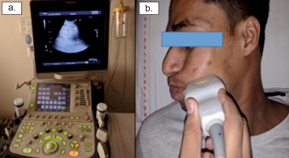
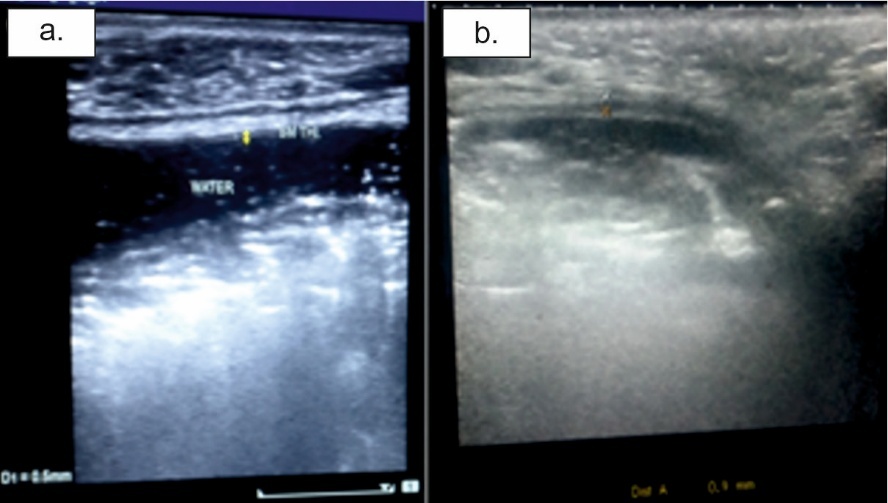
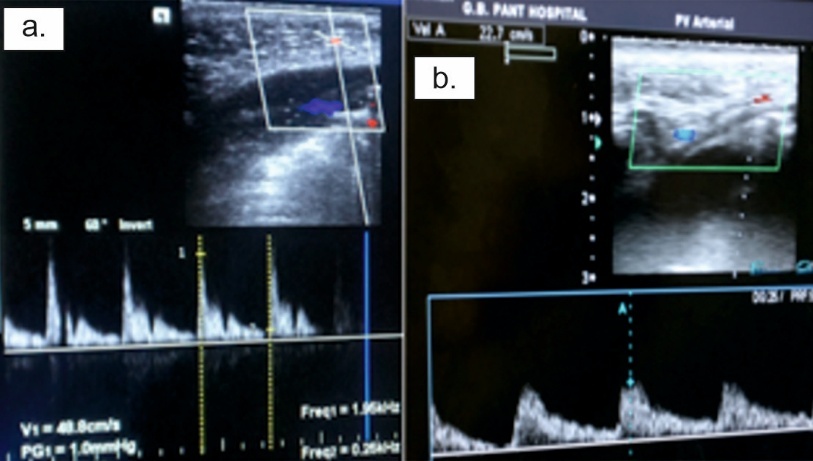
A proficient general radiologist performed ultrasonographic imaging of the patients placed in supine position. The imaging system was a Toshiba high-end ultrasound and color Doppler machine equipped with a 7.5–12 MHz linear probe. ([Figure 1]) The patients were given instructions to retain water in their mouths prior to submucosal thickness measurement in buccal mucosa. Water serves as a coupling medium, enhancing image quality. For precise submucosa thickness measurements, the probe was gently positioned against the surface. For buccal mucosa ultrasonographic imaging, an imaginary line was established between the two reference points: the first point located 1 cm anterior to the masseter muscle's anterior border, and the second point positioned 1 cm posterior to the lip commissure. Submucosal thickness was measured in millimeters along this line bilaterally. Ultrasonographic imaging utilized an extraoral approach with the linear transducer positioned parallel to the mandibular lower border. ([Figure 2]) The mucosal lining appeared as a hyperechoic line, while the submucosa presented as a hypoechoic band supported by hyperechoic muscle layer. This hypoechoic band between the mucosa and muscle layer represented the measured submucosa thickness. The distance between the two hyperechoic lines (mucosa and muscle) was measured using the USG measuring tool ([Figure 3]). Bilateral measurements were recorded, with two scans per patient (right and left sides). Color Doppler assessed vascularity, while spectral Doppler demonstrated blood vessel flow patterns and PSV (Peak systolic value) (Figure 4). PSV measurements were obtained from the most prominent vascular signal within the region of interest, with the highest recorded value used for analysis.
Biopsies were obtained from all patients at representative sites for histopathological examination to confirm clinical diagnosis. Based on histopathological findings, OSMF was classified into stages according to Pindborg JJ and Sirsat SM.[10]
3. Statistical Analysis
Statistical analysis was performed using the statistical software - Statistical Package for Social Sciences, version 19.0, for Windows (SPSS, Chicago, IL). Kruskal-Wallis, t-test, and Pearson Chi-Square tests were applied, and p-values < 0.05 were considered as statistically significant.
4. Results
Our study group included 80 OSMF patients, while the control group comprised 20 subjects. The mean age of the OSMF group was 33.3 years (range: 17–67 years). Group A consisted of 67 males (83.8%) and 13 females (16.2%).
Among Group A participants, 55 patients (68.75%) habitually chewed gutkha, 4 patients (5%) used tobacco, and 1 patient (1.25%) consumed paan masala.
Comparison of mean bilateral submucosal thickness (in mm) and PSV between OSMF and control groups using t-test revealed significantly greater average submucosal thickness in OSMF patients compared to controls (p-value < 0.05). The average PSV was significantly lower in OSMF patients compared to controls, with p-values of 0.001 and 0.004 for right and left sides, respectively. ([Table 1])
Comparison of mean bilateral submucosal thickness and PSV between different histopathological stages in OSMF using Kruskal-Wallis Test demonstrated significant differences in mean submucosal thickness across histopathological stages (p = 0.0001). Mean submucosal thickness increased in the following sequence:
Advanced OSMF > Moderately Advanced OSMF > Early OSMF > Very Early OSMF.
Significant differences were found between mean PSV and histopathological stages (p = 0.0001). Thus, significant differences were observed in average submucosal thickness and Peak Systolic Value among different histopathological stages of OSMF. ([Table 2])
Kruskal-Wallis Test comparison of average bilateral submucosal thickness between different clinical stages in OSMF revealed no significant differences in mean submucosal thickness among clinical stages (p-values 0.256 and 0.323, respectively).
No significant differences in mean bilateral Peak Systolic Values were found among different clinical stages (p-values 0.979 and 0.7176, respectively).
Therefore, no significant differences were established between different clinical stages of OSMF regarding average submucosal thickness and average Peak Systolic Value. ([Table 3])
|
Groups |
RT. B.MT |
LT. B.MT |
PSV RT. B.M |
PSV LT. B.M |
|---|---|---|---|---|
|
Group A (OSF) |
1.299 ± 0.302 |
1.321 ± 0.320 |
30.787 ± 5.710 |
31.691 ± 6.590 |
|
Group B (Control) |
0.560 ± 0.094 |
0.550 ± 0.089 |
41.205 ± 11.471 |
39.725 ± 10.837 |
|
t-test (p value) |
0.0001 |
0.0001 |
0.001 |
0.004 |
|
Histopathological stage |
RT. B.MT |
LT. B.MT |
PSV RT. B.M |
PSV LT. B.M |
|---|---|---|---|---|
|
Very early OSF |
0.725 ± 0.05 |
0.70 ± 0.50 |
37 ± 3.92 |
39.9 ± 2.69 |
|
Early OSF |
0.952 ± 0.156 |
0.957 ± 0.163 |
35.257 ± 4.820 |
37.329 ± 5.81 |
|
Moderately advanced OSF |
1.457 ± 0.139 |
1.488 ± 0.149 |
28.580 ± 4.772 |
28.920 ± 4.931 |
|
Advanced OSF |
1.657 ± 0.126 |
1.725 ± 0.096 |
29.250 ± 5.582 |
29.225 ± 7.386 |
|
Kruskal-Wallis Test (p- value) |
0.0001 |
0.0001 |
0.0001 |
0.0001 |
|
Clinical staging |
RT. B.MT |
LT. B.MT |
PSV RT. B.M |
PSV LT. B.M |
|---|---|---|---|---|
|
Stage 1 |
1.246 ± 0.325 |
1.268 ± 0.350 |
30.805 ± 5.835 |
32.432 ± 6.902 |
|
Stage 2 |
1.332 ± 0.281 |
1.355 ± 0.294 |
30.800 ± 5.647 |
31.117 ± 6.549 |
|
Stage 3 |
1.500 ± 0.173 |
1.533 ± 0.116 |
30.400 ± 7.286 |
30.200 ± 1.312 |
|
Kruskal-Wallis Test (p-value) |
0.256 |
0.323 |
0.979 |
0.717 |
5. Discussion
OSMF fibrosis represents a dynamic process requiring periodic disease activity monitoring to evaluate responses to habit cessation or medical interventions. Biopsy, as a minor surgical procedure, causes patient discomfort and is time-consuming. Additionally, biopsy has limitations in diffuse diseases like OSMF, where single-site tissue sampling may not represent the true disease severity extent. Furthermore, biopsy site scarring and increased fibrosis may occur. Ultrasonography is particularly appropriate for head and neck superficial structure imaging, providing both qualitative and quantitative assessments. It can detect lesion characteristics, dimensions, distance from skin surfaces, and proximity to mucosal structures.
Shah et al., noted that gutkha chewing produced OSMF more rapidly compared to raw areca nut and other products.[11] Increased chewing habit frequency was correlated with disease severity due to indirect pathogenic relationships, where normal oral mucosa experiences greater areca nut exposure. The areca nut acts as a chronic irritant and causes mechanical trauma, while slaked lime, a gutkha ingredient, causes chemical burns which leads to microhemorrhage. Both of these result in increased fibrosis due to fibroblast permeation.[12] Combined smoking and smokeless tobacco use was observed in 17 patients (21.25%), with no patients using smoking tobacco exclusively. This indicates that smoking alone does not contribute to OSMF, consistent with Shah et al., findings.[11] Smokeless tobacco product chewing habits represent more predominant OSMF etiological factors than smoking or combined tobacco habits.
Increased habit duration was associated with reduced interincisal opening in OSMF cases, consistent with studies by Pandya et al, Devathambhi et al.[13][14] The increased OSMF severity due to prolonged habit duration can be attributed to chronic contact between areca nut mixture and oral mucosa, causing repeated mechanical and chemical trauma leading to increased connective tissue fibrosis and reduced interincisal opening.
Control group average bilateral submucosal thickness measured 0.560 ± 0.094 mm and 0.550 ± 0.089 mm, respectively. This observation aligns with Devathambhi et al., study showing normal patient submucosal thickness of 0.505 ± 0.052mm.[14] Control group mean bilateral Peak Systolic Values measured 41.205 ± 11.471 cm/sec and 39.725 ± 10.837 cm/sec, respectively, consistent with Gandhi et al., study showing buccal mucosa PSV range of 43.87 ± 20.95 cm/sec.[15]
In the OSMF group, average bilateral submucosal thickness measured 1.299±0.302 mm and 1.321 ± 0.320 mm, respectively, with no significant bilateral differences. OSMF group average submucosal thickness significantly exceeded control group measurements (p-value < 0.05), consistent with studies by Rangaiah et al, Devathambhi et al.[12][14] Since OSMF is characterized by increased lamina propria and submucosal fibrosis, ultrasonographic submucosal layer thickness increases were observed. Therefore, OSMF causes significant submucosa thickness increases that can be clearly evaluated ultrasonographically.
PSV in OSMF patients was significantly reduced as compared to controls (p-value < 0.05), with no significant bilateral differences. This finding is consistent with Manjunath et al., study demonstrating similar vascularity reduction with disease progression using Color Doppler USG.[7]
This study found significant increases in average buccal mucosa thickness and significant reductions in peak systolic value (PSV) with advancing histopathological and clinical OSMF stages (p-value < 0.05). Therefore, as OSMF severity increases, submucosal thickness increases due to increased fibrosis. PSV also decreases with OSMF advancement due to reduced vascularity with disease progression. Study limitations included USG operator dependence, single-radiologist evaluation, lack of blinding, small control group size and unassessed inter-observer and intra-observer reliability. These should be addressed in future research.
6. Conclusion
Since OSMF is a diffuse disease, performing biopsies from all four sites in all patients for histopathological evaluation may not be feasible or ethical. Additionally, biopsies may not be completely representative of disease severity in diffuse conditions. Although biopsy can be considered the diagnostic gold standard for OSMF, its role as a gold standard for measuring disease severity across the entire buccal mucosa is questionable.
USG evaluation in OSMF demonstrated that when real-time US is combined with color Doppler US, it provides a valuable non-invasive tool for oral subcutaneous tissue assessment, thereby avoiding invasive, time-consuming biopsies.
7. Authors Contributions
Sunita Gupta: Conceptualization, scientific advisor. Puneet Vats: Data curation, investigation. Poonam Narang: Resources. Vineeta Vijay Batra: Formal analysis, resources. Sujoy Ghosh: Conceptualization, formal analysis. Kavita Vishal Khari: Scientific advisor, writing – review editing.
8. Source of Funding
None.
9. Conflict of Interest
None.
10. Ethical Approval
The following study was approved by the Institutional Ethics Committee of Maulana Azad Institute of Dental Sciences, New Delhi, India with (Ref.No. MAIDS/Ethical Committee/2016, dated 30th October 2016.
References
- Arakeri G, Rai KK, Hunasgi S, Merkx MAW, Gao S, Brennan PA. Oral submucous fibrosis: An update on current theories of pathogenesis. J Oral Pathol Med. 2017;46(6):406-412. [Google Scholar] [Crossref]
- Passi D, Bhanot P, Kacker D, Chahal D, Atri M, Panwar Y. Oral submucous fibrosis: Newer proposed classification with critical updates in pathogenesis and management strategies. Natl J Maxillofac Surg. 2017;8(2):89-94. [Google Scholar] [Crossref]
- Ekanayaka RP, Tilakaratne WM. Oral submucous fibrosis: review on mechanisms of malignant transformation. Oral Surg Oral Med Oral Pathol Oral Radiol. 2016;122(2):192-199. [Google Scholar] [Crossref]
- Caglayan F, Bayrakdar IS. The Intraoral Ultrasonography in Dentistry. Niger J Clin Pract. 2018;21(2):125-133. [Google Scholar] [Crossref]
- Benson BW, Flint DJ, Liang H, Opatowsky MJ. Advances in diagnostic imaging for pathologic conditions of the jaws. Head Neck Pathol. 2014;8(4):383-s91. [Google Scholar] [Crossref]
- Joshi PS, Pol J, Sudesh AS. Ultrasonography - A diagnostic modality for oral and maxillofacial diseases. Contemp Clin Dent. 2014;5(3):345-351. [Google Scholar] [Crossref]
- Manjunath K, Rajaram PC, Saraswathi TR, Sivapathasundharam B, Sabarinath B, Koteeswaran D, et al. Evaluation of oral submucous fibrosis using ultrasonographic technique: a new diagnostic tool. Indian J Dent Res. 2011;22(4):530-536. [Google Scholar] [Crossref]
- Reddy V, Wanjari PV, Banda NR, Reddy PSN. Oral Submucous Fibrosis: Correlation of Clinical Grading to various habit factors. Int J Dent Clin. 2011;3(1). [Google Scholar] https://scispace.com/papers/oral-submucous-fibrosis-correlation-of-clinical-grading-to-4mn8vh1ljw
- Haider SM, Merchant AT, Fikree FF, Rahbar MH. Clinical and functional staging of oral submucous fibrosis. Br J Oral Maxillofac Surg. 2000;38(1):12-15. [Google Scholar] [Crossref]
- Pindborg JJ, Murti PR, Bhonsle RB, Gupta PC, Daftary DK, Mehta FS. Oral submucous fibrosis as a precancerous condition. Scand J Dent Res. 1984;92(3):224-229. [Google Scholar] [Crossref]
- Shah N, Sharma PP. Role of chewing and smoking habits in the etiology of oral submucous fibrosis (OSF): a case-control study. J Oral Pathol Med. 1998;27(10):475-479. [Google Scholar] [Crossref]
- Rangaiah P, Annigeri RG, Lingappa A. The Changes are Incorporated in Red Color Font Transcutaneous Ultrasonographic Assessment of Oral Submucous Fibrosis : A Preliminary Study. Int J Oral Med Sci. 2010;9(2):137-147. [Google Scholar] [Crossref]
- Pandya S, Chaudhary AK, Singh M, Singh M, Mehrotra R. Correlation of histopathological diagnosis with habits and clinical findings in oral submucous fibrosis. Head Neck Oncol. 2009;1(1):10. [Google Scholar] [Crossref]
- Devathambi JR, Aswath N. Ultrasonographic evaluation of oral submucous fibrosis and masseteric hypertrophy. J Clin Imaging Sci. 2013;3(Suppl 1):12. [Google Scholar] [Crossref]
- Gandhi R, Bhowate R, Nayyar AS, Gandhi S, Dongarwar G. Color Doppler-ultrasonography in oral squamous cell carcinoma: Making ultrasonography more meaningful. Adv Biomed Res. 2016;5(1):29. [Google Scholar] [Crossref]
Article Metrics
- Visibility 122 Views
- Downloads 125 Views
- DOI 10.18231/j.jicd.77609.1792216918
-
CrossMark

- Citation
- Received Date July 23, 2026
- Accepted Date August 27, 2026
- Publication Date October 17, 2026