Journal of International College of Dentists
Journal of International College of Dentists (JICD) is an open-access, peer-reviewed, Quarterly journal publishing under the auspices of the International College of Dentists. As soon as the Article is accepted for Publication, it will be placed in “Ahead of Print” on the journal page with the aim of rapid and better knowledge dissemination. In addition, the journal allows free access (Open Access) to its contents, which is likely to attract more readers and citations to articles published in journal. Manuscripts must be prepared under the “Uniform requirements” of the ...
A retrospective cephalometric study on upper airway spaces in different facial types in Class II malocclusion

Abstract
Introduction: Craniofacial growth pattern has been correlated with variations in size of the upper airway spaces. The objective of this study was to evaluate the nasopharyngeal, oropharyngeal, and hypopharyngeal airway spaces variations according to the craniofacial growth pattern, by comparing brachyfacial, mesofacial, and dolichofacial in Angle Class II individuals.
Materials and Methods: To measure the spaces, 90 lateral cephalograms were used and divided into 3 groups per the craniofacial growth pattern, determined by the Tweed cephalometry angular measurements: FMA and Y-axis. To evaluate the airways, sleep apnea cephalometry was used, containing 19 points that compose 13 factors. Three groups were compared relative to each of the sleep apnea cephalometry measurements.
Results: Statistically significant difference was verified among the groups for the measurement of the median posterior-palatal space, Tongue length, Superior pharyngeal space and posterior airway space with the difference being pointed out between the brachyfacial, mesofacial and dolichofacial groups. For the other measurements, there was no statistically significant difference.
Conclusion: The study concluded that skeletal Class II dolichofacial individuals showed reduced oropharyngeal space with decreased median posterior palatal measurement, while brachyfacial individuals exhibited increased hypopharyngeal space due to greater tongue length and posterior airway dimensions.
1. Introduction
The upper airway consists of three main regions: the nasopharynx, oropharynx, and hypopharynx. The dimensions of the pharyngeal airway space are primarily influenced by the growth and relative size of the soft tissues surrounding the dentofacial structures.[1][2] Adequate upper airway development facilitates normal nasal breathing and plays an important role in craniofacial growth and development.[1][2]
Obstruction of the upper airway may occur due to various morphological, physiological, or pathological factors, including adenoid and tonsillar hypertrophy, chronic or allergic rhinitis, environmental irritants, infections, congenital nasal abnormalities, nasal trauma, polyps, and tumors. These conditions may disrupt normal airway function and contribute to the development of mouth-breathing patterns.[3]
Upper airway dimensions are also influenced by the underlying facial skeletal pattern, particularly the anteroposterior relationship between the maxilla and mandible, which significantly affects available airway space.[1] Previous studies have investigated airway changes associated with orthodontic treatment, orthognathic surgery, and obstructive sleep apnea conditions.[4] However, limited evidence is available regarding the relationship between airway dimensions, facial growth patterns, and individuals with Angle Class II malocclusion.
Understanding these variations may provide valuable information for orthodontic diagnosis and treatment planning.[5]
Therefore, the present study aimed to assess and compare the nasopharyngeal, oropharyngeal, and hypopharyngeal airway dimensions among brachyfacial, mesofacial, and dolichofacial individuals with Angle Class II malocclusion.
2. Materials and Methods
The study sample consisted of 90 digital lateral cephalometric radiographs of individuals undergoing orthodontic treatment in National Dental College having complete permanent dentition and skeletal Class II relationship, determined according to Steiner’s ANB angle analysis.[6]
The sample was divided into three groups based on facial type: Group I (n = 30): Mesofacial. Group II (n = 30): Dolicofacial. Group III (n = 30): Brachyfacial.
Radiographs were excluded if they showed poor visualization of anatomical structures, history of palatal tonsil or pharyngeal tonsil surgery, previous orthodontic or orthognathic treatment, dental extractions, or congenital dental agenesis.
All lateral cephalograms were obtained using the same radiographic equipment (Carestream CS 8100 unit). Cephalometric analysis was performed manually by same operator.
Facial growth patterns were determined using Tweed’s Frankfort mandibular plane angle (FMA) and the Y-axis measurements.[7] The radiographs were randomly selected and classified according to these parameters.
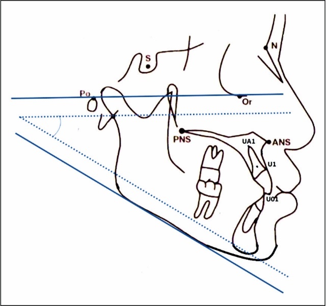
The FMA angle was calculated between the mandibular plane and the Frankfort horizontal plane (Po-Or), with a reference value of 25°. An FMA value greater than 30° indicated a vertical growth pattern (dolichofacial), whereas values below 20° represented a horizontal growth pattern (brachyfacial). ([Figure 1])
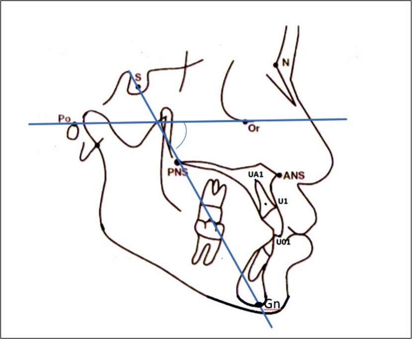
The Y-axis, also known as the facial growth angle, was determined by the intersection of the Sella-Gnathion line and the Frankfort horizontal plane. The average reference value was considered 59°. Increased Y-axis values indicated a vertical growth tendency, while decreased values suggested horizontal growth.[8] ([Figure 2])
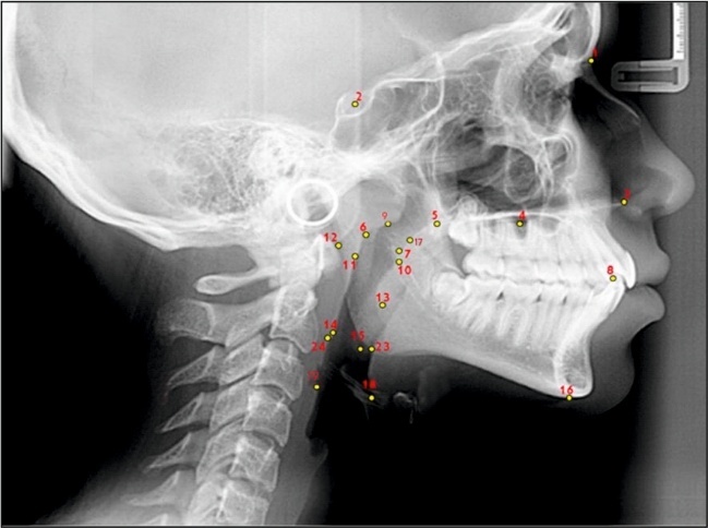
30 subjects were included in each facial growth group: mesofacial, brachyfacial, and dolichofacial. To evaluate the upper airways, sleep apnea cephalometry, was used, containing 19 points that compose 13 factors. ([Table 1]), ([Figure 3])
These are the landmarks measured on the lateral cephalogram. ([Table 2]) ([Figure 4],[5])
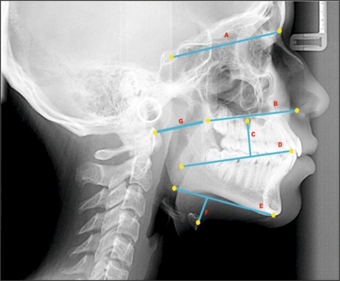
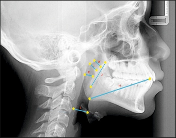
|
S.No. |
Cephalometric Points |
Definition |
|---|---|---|
|
1 |
N |
Nasion |
|
2 |
S |
Sella |
|
3 |
ANS |
Anterior nasal spine |
|
4 |
DI |
Tongue dorsum |
|
5 |
PNS |
Posterior nasal spine |
|
6 |
PP2 |
Posterior palate 2 |
|
7 |
Pp2’ |
PP2 projection to posterior pharynx wall |
|
8 |
Tt |
Tongue tip |
|
9 |
PPFS |
Posterior nasopharynx wall |
|
10 |
PAFM |
Anterior medium pharynx wall |
|
11 |
PPFM |
Posterior medium pharynx wall |
|
12 |
At |
Atlas vertebra |
|
13 |
P |
Lowest point of the soft palate |
|
14 |
Bgo |
Me-Goc projection to post. laryngopharynx Wall |
|
15 |
Goc |
Cephalometric gonion |
|
16 |
Me |
Menton |
|
17 |
PP1 |
Posterior palate 1 |
|
18 |
H’ |
Hyoid Projection to ant. Laryngopharynx Wall |
|
19 |
C3’ |
3rd Cervical vertebra |
|
S.No. |
Cephalometric landmarks |
Definition |
|---|---|---|
|
1 |
(N-S) |
Anterior base of cranium |
|
2 |
(ANS-PNS) |
Maxillary length |
|
3 |
TGH |
Tongue-Height |
|
4 |
TGL |
Tongue-Length |
|
5 |
(Me-Goc) |
Mandibular length |
|
6 |
(MP(Me-Goc)/H) |
Hyoid-Mandibular plane distance |
|
7 |
(At-PNS) |
Atlas-Maxilla distance |
|
8 |
(PPFS-PP1) |
Superior pharyngeal space |
|
9 |
(Pp2-Pp2’) |
Median posterior palatal space |
|
10 |
(PNS-P) |
Soft palate length |
|
11 |
(PPFM-PAFM) |
Middle pharyngeal space |
|
12 |
(C3’-H’) |
Inferior pharyngeal space |
|
13 |
(Bgo-Tt) |
Posterior airway space |
|
Particulars |
Mesofacial (Mean ± SD) |
Dolicofacial (Mean ± SD) |
Brachyfacial (Mean ± SD) |
p-value |
|---|---|---|---|---|
|
N-S |
67.30 ± 3.82 |
66.20 ± 3.85 |
67.13 ± 3.45 |
0.468 |
|
ANS-PNS |
51.37 ± 4.17 |
51.27 ± 4.14 |
52.90 ± 4.51 |
0.258 |
|
TGH |
19.70 ± 2.59 |
20.83 ± 2.04 |
19.37 ± 2.66 |
0.057 |
|
TGL |
65.27 ± 4.56 |
67.83 ± 4.65 |
68.33 ± 5.50 |
0.040 |
|
Me-GOC |
65.90 ± 4.08 |
64.60 ± 4.02 |
66.83 ± 4.95 |
0.145 |
|
MP-H |
12.10 ± 2.31 |
11.80 ± 3.01 |
12.23 ± 2.47 |
0.806 |
|
At-PNS |
36.40 ± 3.40 |
35.53 ± 3.81 |
37.33 ± 4.32 |
0.202 |
|
PPFS-PP1 |
13.20 ± 2.68 |
10.87 ± 2.50 |
12.73 ± 2.30 |
0.001 |
|
PP2-PP2’ |
11.50 ± 2.32 |
9.87 ± 1.94 |
11.37 ± 2.46 |
0.010 |
|
PNS-P |
30.70 ± 3.28 |
31.37 ± 3.51 |
30.63 ± 4.85 |
0.729 |
|
PPFM-PAFM |
9.23 ± 2.01 |
9.30 ± 1.78 |
10.33 ± 2.34 |
0.073 |
|
H’-C3’ |
10.13 ± 3.44 |
11.33 ± 3.12 |
11.53 ± 3.97 |
0.257 |
|
Bgo-Tt |
75.17 ± 5.88 |
75.60 ± 6.72 |
80.20 ± 6.30 |
0.004 |
3. Results
When we compared mesofacial, dolicofacial and branchyfacial facial pattern with each other, the mean difference found to be significant in case of:([Table 3]) TGL (Tongue length) (p = 0.04). PPFS-PP1(Superior pharyngeal space) (p = 0.001). PP2-PP2’(Median posterior palatal space) (p = 0.010). Bgo-Tt (Posterior airway space) (p = 0.004). While the other parameters are found to be non-signficant in all cases.
4. Discussion
The present study emphasizes the role of conventional orthodontic cephalometric analysis in assessing upper airway anatomical variations associated with respiratory disorders. Lateral cephalometry is considered a reliable and accessible diagnostic method due to its reproducibility, cost-effectiveness, and low radiation exposure. Previous studies have extensively explored the relationship between craniofacial morphology and airway dimensions to identify potential airway-related risk factors.[9]
Although CBCT and other three-dimensional imaging techniques provide comprehensive evaluation of airway morphology, including volume and cross-sectional area analysis, their routine clinical application is limited due to increased radiation exposure and cost considerations. The present study was limited by the inability to evaluate airway volume using two-dimensional lateral cephalograms.[10] However, potential errors related to superimposition in cephalometric analysis were minimized as no superimposition procedures were performed.
Previous studies have assessed upper airway dimensions using lateral cephalometry and demonstrated associations between airway morphology, skeletal pattern, and facial growth characteristics. Ceylan and Oktay reported that sagittal and vertical skeletal discrepancies significantly influence pharyngeal airway dimensions, with reduced airway space in unfavorable skeletal relationships.[5] Longitudinal studies have further supported the reliability of lateral cephalometry for evaluating nasopharyngeal changes among different growth patterns.[11] In the present study, sleep apnea cephalometry was used due to its validated and standardized airway measurements.[8]
The present study demonstrated a significant reduction in superior pharyngeal space and median posterior palatal space in dolichofacial individuals compared with brachyfacial and mesofacial groups. This may be attributed to the vertical growth pattern in dolichofacial subjects, where mandibular downward and backward rotation results in reduced oropharyngeal dimensions. The tongue base follows this rotational pattern, positioning posteriorly and causing retrusion of the soft palate. Hochban et al., reported that mandibular retrusion and clockwise rotation may contribute to posterior displacement of the tongue and soft palate, increasing airway narrowing risk.[6]
The greatest airway alterations in individuals with OSAS are commonly observed in the oropharyngeal region, particularly involving reduction of the median posterior palatal space.[8] Additionally, the tongue base follows the mandibular rotational pattern, moving downward and backward, which may position the soft palate more posteriorly and contribute to narrowing of the median posterior palatal space.[12]
Significant differences were observed in tongue length and posterior airway space, with brachyfacial individuals showing greater values compared with mesofacial and dolichofacial groups.[13] This may be due to the horizontal growth pattern, where forward and upward mandibular rotation increases tongue positioning space and posterior pharyngeal dimensions.[14][15] Muto et al., reported that mandibular position and growth direction significantly influence tongue posture and pharyngeal airway dimensions.[7]
Upper airway obstruction may result in compensatory mouth breathing, leading to altered orofacial functions such as lip incompetence, lowered tongue posture, tongue thrusting, and muscular imbalance, which may influence swallowing, mastication, speech, and occlusal stability. A limitation of this study was the use of two-dimensional cephalometry; future studies using CBCT may provide better airway evaluation while considering radiation exposure and clinical justification.
5. Conclusion
Under the conditions of this study and considering the results, it is concluded that there was significant reduction in Oropharyngeal space in skeletal Class II dolichofacial individuals, characterized by reduction in the median posterior palatal measurement.
There is increase in hypopharyngeal space in skeletal class II brachyfacial individuals that is characterized by increase in tongue length and posterior airway space.
6. Authors Contributions
Kashish Goyal: Conceptualization, data curation, formal analysis, investigation, resources, software, visualization, writing – original draft. Vinay Dua: Conceptualization, formal analysis, investigation, scientific advisor. Harvinder Singh: Formal analysis, methodology, scientific advisor, visualization. Yenika Manchanda: Formal analysis, methodology, scientific advisor. Amanjot Kaur: Data curation, scientific advisor, visualization.
7. Source of Funding
None.
8. Conflict of Interest
None.
References
- Lopatienė K, Dabkutė A, Juškevičiūtė V. Vertical and sagittal morphology of the facial skeleton and the pharyngeal airway. Stomatologija. 2016;18(1):21-25. [Google Scholar]
- Ghoneima A, AlBarakati S, Jiang F, Kula K, Wasfy T. Computational fluid dynamics analysis of the upper airway after rapid maxillary expansion: a case report. Prog Orthod. 2015;16(1):10. [Google Scholar] [Crossref]
- Claudino LV, Mattos CT, Ruellas AC, Sant' Anna EF. Pharyngeal airway characterization in adolescents related to facial skeletal pattern: a preliminary study. Am J Orthod Dentofacial Orthop. 2013;143(6):799-809. [Google Scholar] [Crossref]
- Borges Pde T, Silva BB, Moita Neto JM, Borges NE, Li LM. Cephalometric and anthropometric data of obstructive apnea in different age groups. Braz J Otorhinolaryngol. 2015;81(1):79-84. [Google Scholar] [Crossref]
- Ceylan I, Oktay H. A study on the pharyngeal size in different skeletal patterns. Am J Orthod Dentofacial Orthop. 1995;108(1):69-75. [Google Scholar] [Crossref]
- Hochban W, Brandenburg U, Peter JH. Surgical treatment of obstructive sleep apnea by maxillomandibular advancement. Sleep. 1994;17(7):624-629. [Google Scholar] [Crossref]
- Muto T, Yamazaki A, Takeda S. A cephalometric evaluation of the pharyngeal airway space in patients with mandibular retrognathia and prognathia, and normal subjects. Int J Oral Maxillofac Surg. 2008;37(3):228-231. [Google Scholar] [Crossref]
- Park JE, Gray S, Bennani H, Antoun JS, Farella M. Morphometric growth changes of the nasopharyngeal space in subjects with different vertical craniofacial features. Am J Orthod Dentofacial Orthop. 2016;150(3):451-458. [Google Scholar] [Crossref]
- Saghaie S, Ghaffari R. Effect of image compression of direct digital lateral cephalograms on the identification of cephalometric points. Dent Res J (Isfahan). 2014;11(1):27-31. [Google Scholar]
- Tweed CH. Was the development of the diagnostic facial triangle as an accurate analysis based on fact or fancy? Am J Orthod. 1962;48(11):823-840. [Google Scholar] [Crossref]
- Maschtakow PS, Tanaka JL, da Rocha JC, Giannas LC, de Moraes ME, Costa CB, et al. Cephalometric analysis for the diagnosis of sleep apnea: a comparative study between reference values and measurements obtained for Brazilian subjects. Dental Press J Orthod. 2013;18(3):143-149. [Google Scholar] [Crossref]
- Aboudara C, Nielsen I, Huang JC, Maki K, Miller AJ, Hatcher D. Comparison of airway space with conventional lateral headfilms and 3-dimensional reconstruction from cone-beam computed tomography. Am J Orthod Dentofacial Orthop. 2009;135(4):468-479. [Google Scholar] [Crossref]
- Tsunori M, Mashita M, Kasai K. Relationship between facial types and tooth and bone characteristics of the mandible obtained by CT scanning. Angle Orthod. 1998;68(6):557-562. [Google Scholar] [Crossref]
- Silva VG, Pinheiro LA, Silveira PL, Duarte AS, Faria AC, Carvalho EG, et al. Correlation between cephalometric data and severity of sleep apnea. Braz J Otorhinolaryngol. 2014;80(3):191-195. [Google Scholar] [Crossref]
- Cho HW, Kim IK, Cho HY, Seo JH, Lee DH, Park SH. Retrospective study of changes in pharyngeal airway space and position of hyoid bone after mandibular setback surgery by cephalometric analysis. Maxillofac Plast Reconstr Surg. 2015;37(1):38. [Google Scholar] [Crossref]
Article Metrics
- Visibility 132 Views
- Downloads 138 Views
- DOI 10.18231/j.jicd.30832.1792217141
-
CrossMark

- Citation
- Received Date July 03, 2026
- Accepted Date August 29, 2026
- Publication Date October 17, 2026