Panacea Journal of Medical Sciences
Panacea Journal of Medical Sciences (PJMS) open access, peer-reviewed triannually journal publishing since 2011 and is published under auspices of the “NKP Salve Institute of Medical Sciences and Research Centre”. With the aim of faster and better dissemination of knowledge, we will be publishing the article ‘Ahead of Print’ immediately on acceptance. In addition, the journal would allow free access (Open Access) to its contents, which is likely to attract more readers and citations to articles published in PJMS.Manuscripts must be prepared in accordance with “Uniform requiremen...

Precision in practice: Comparative evaluation of cytomorphological findings, acid-fast bacilli staining and cartridge-based nucleic acid amplification test for diagnosing suspected cervical tubercular lymphadenopathy
- Author Details:
-
 Anushree C N ,
-
Shaista Choudhary ,
- Seema Sanghamitra Maharana ,
- Bhavya S Nair * ,
- Krisha M ,
- Akshay Raju
Introduction
Before the COVID-19 pandemic, Tuberculosis (TB) caused more deaths than any other single infectious agent, including HIV/AIDS.[1] As of 2021, the number of TB patients notified has increased by 19% compared to 2020. [2]
Tuberculosis (TB) is an ancient bacterial disease affecting humans, caused by Mycobacterium tuberculosis. It manifests in two main types: pulmonary TB (PTB) and extrapulmonary TB (EPTB). Extrapulmonary TB occurs when the infection affects organs outside the lungs, with the lymph nodes being the most frequently affected site. [3] EPTB cases constitute approximately 15-20% of all TB cases. [4]
The most common manifestation of mycobacterial infections is cervical lymphadenitis, [5] and TB of the head and neck can be difficult to diagnose due to a large number of smear-negative cases. This often leads to positive cases being missed, which increases the burden of TB. [6]
Conventional methods for diagnosing EPTB include fine-needle aspiration cytology of lymph nodes and demonstrating acid-fast bacilli (AFB) using Ziehl-Neelson (ZN) staining. [7] Fine-needle aspiration cytology of lymph nodes can be presumptively diagnosed with tuberculous lymphadenitis morphologically. [8] The cartridge-based nucleic acid amplification test (CBNAAT) is a real-time polymerase chain reaction (PCR) assay designed to swiftly detect the presence of Mycobacterium tuberculosis and rifampicin resistance in biological samples, providing results within a two-hour timeframe. Acknowledged by the World Health Organization (WHO) in December 2010, this technology stands as a pivotal advancement in global tuberculosis (TB) control efforts. [9]
Materials and Methods
The prospective study was done at DR B. R Ambedkar Medical College & Hospital, Bangalore from October 2021 to January 2023. The study consists of clinically suspected cervical tubercular lymphadenopathy patients referred from various departments of this hospital for FNAC.
Inclusion criteria
Patients aged above 18 years and below 65 years who are clinically suspected with cervical tubercular lymphadenopathy.
Exclusion criteria
Individuals with a confirmed history of tuberculosis.
Patients currently undergoing anti-tubercular drug treatment.
Cases where aspirates or samples were insufficient for a cytopathological diagnosis.
Patients who declined to provide consent for participation.
Study procedure
Prior to conducting Fine-Needle Aspiration Cytology (FNAC), the patients were provided with an informed consent form in the language of their choice. Their clinical history and relevant investigations were thoroughly reviewed. During the Fine Needle Aspiration Cytology (FNAC) procedure, a 22-gauge needle, coupled with a 10 ml syringe, was employed to ensure the collection of sufficient and representative samples. Adequate smears were made and immediately fixed and prepared for cytomorphological analysis using Hematoxylin and Eosin (H&E) and Papanicolaou stain (PAP). One smear was air-dried and stained for acid-fast bacilli while the remaining aspirate was rinsed into a Falcon tube with 5 ml of normal saline and processed for CB-NAAT testing. The procedures were executed in adherence to the operator manual specifications outlined by the Central TB Division, Government of India, for the Gene Xpert system. The assay sample reagent, comprising a mixture of sodium hydroxide and isopropanol in a 2:1 ratio, was meticulously introduced to the sample. Following a 15-minute incubation period at room temperature with periodic agitation, 3ml of the processed sample was meticulously loaded into the cartridge and seamlessly integrated into the CB-NAAT instrument module. The subsequent assay steps and conclusive results were automatically presented on the Gene Xpert monitor approximately 1 hour and 50 minutes after initiation.[10]
Ethics
The study was conducted only after receiving approval from the Institutional Ethics Committee (IEC), with reference number 158.
Statistical analysis
Microsoft Word and Excel generated graphs, tables, etc. Data was coded and entered into a statistical package for social science (SPSS) software version 20.00 and analyzed.
Results
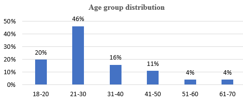
A total of 102 cases underwent FNAC based on clinical and radiological findings. Females were the majority ([Table 1]) and the most common age group was 21-30 [[Figure 1]].
|
Gender |
Number of cases |
|
Male |
42 |
|
Female |
60 |
Upon examination, all cases showed lymph node enlargement. Of these, 63 cases were discrete, and 39 were matted. Most cases were found in level V, as shown in [[Table 2]].
|
Level of lymph node |
No. of cases |
|
I |
13 |
|
II |
5 |
|
III |
14 |
|
IV |
26 |
|
V |
44 |
During aspiration, most cases showed blood mixed with aspirate. The CBNAAT and AFB tests were more positive in the caseous aspirate. [[Table 3]]
|
Types of aspirates |
Number |
No of CBNAAT positive cases |
No AFB-positive cases |
|
Blood mixed |
65 |
23 |
6 |
|
Purulent |
29 |
24 |
8 |
|
Caseous |
8 |
7 |
5 |
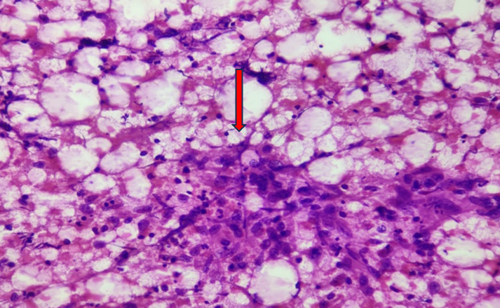
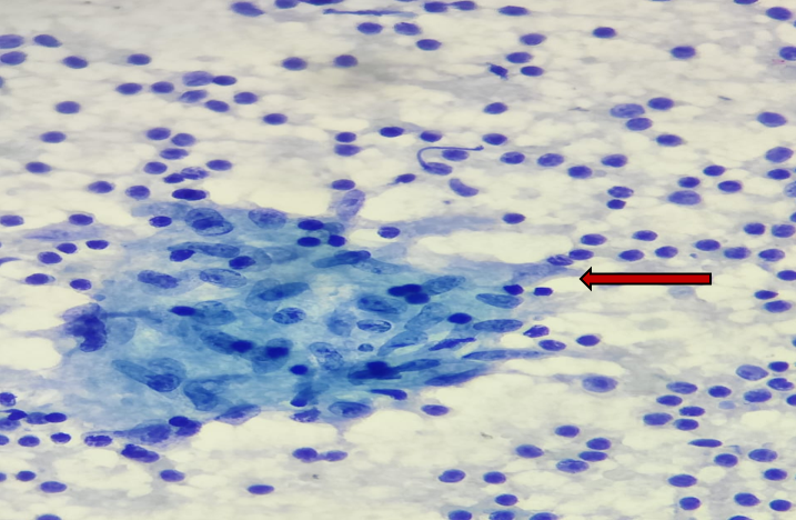
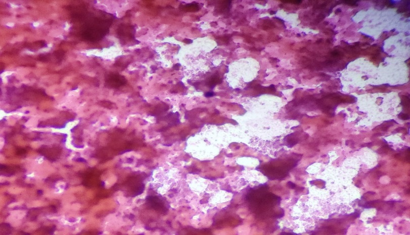
Among cytomorphological findings of 102 cases [[Table 4]], 46 cases showed granulomatous lymphadenitis with necrosis [[Figure 2]],12 cases showed granulomatous lymphadenitis without necrosis [[Figure 3]] and 3 cases showed necrotizing lymphadenitis [[Figure 4]].
|
Cytomorphological findings |
Number of cases |
Percentage |
No of CBNAAT positive cases |
No AFB Positive cases |
|
Granulomatous lymphadenitis with necrosis |
46 |
45.09 % |
39 |
14 |
|
Granulomatous lymphadenitis without necrosis |
12 |
11.76 % |
8 |
3 |
|
Necrotizing lymphadenitis |
3 |
2.94 % |
3 |
2 |
|
Reactive lymphadenitis |
25 |
24.5% |
2 |
0 |
|
Lymphoproliferative disorder |
5 |
4.90% |
0 |
0 |
|
Mets |
5 |
4.90% |
0 |
0 |
|
Suppurative inflammatory disease |
4 |
3.92% |
2 |
0 |
|
Chronic lymphadenitis |
2 |
1.96% |
0 |
0 |
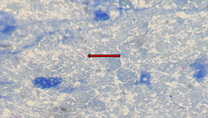
In this study, among 102 cases 54 cases showed CBNAAT positive, and 19 cases showed AFB positive [[Figure 5]].
|
Study |
Period |
Age group |
% of cases |
Females |
|
Present study |
Oct 2021 – Jan 2023 |
21-30 |
46% |
58.82% |
|
Mulualem et al |
May-Sept 2013 |
16-30 |
58% |
53.1% |
|
Bryan Rock et al[11] |
Jan1993-Dec 2003 |
15-24 |
43% |
54% |
|
Adhikary et al[12] |
Mar 2021- Feb 2022 |
21 - 30 |
27.85 % |
59.49 % |
|
Study |
Study Period |
Sensitivity |
Specificity |
|
Present study |
October 2021-January 2023 |
81.97% |
90.2% |
|
Arpitha et al[13] |
July 2019- June 2020 |
80% |
86% |
|
Suwarna B. Patil et al[14] |
January 2019 - December 2019 |
55.5 % |
83.80% |
|
Lavanya et al[15] |
April 2018 to March 2019 |
28.75% |
88.7% |
|
Komanapalli SK et al[16] |
April 2017- March 2018 |
84.25 % |
86.71% |
In total, CBNAAT exhibited positivity in 54 cases, while AFB demonstrated positivity in 19 cases. In comparison to FNAC, CBNAAT displayed a sensitivity of 81.97%, a specificity of 90.2%, a positive predictive value of 92.6%, and a negative predictive value of 77.1%. In contrast, AFB exhibited a sensitivity of 31.15%, a specificity of 100%, a positive predictive value of 100%, and a negative predictive value of 49.4%.
Discussion
This prospective study, conducted at a tertiary care hospital, aimed to diagnose cervical tubercular lymphadenitis by comparing FNAC with AFB staining and CBNAAT. FNAC, recognized as the primary diagnostic test, holds significance in detecting tubercular lymphadenitis.[17]
In this study, we have included only cervical lymph nodes as it is the most common site of EPTB.[3]
This present study shows the greatest number of cases affected in the 21-30 age group with female preponderance. This is similar to studies conducted by Mulualem et al, Bryan Rock et al and Adhikary et al[18], [11], [12] [[Table 5]].
All 102 cases showed lymph node enlargement and the highest number of patients presented with discrete swelling (61.76 %) and the majority of the lymph nodes were seen in level V (43.14 %). These findings are comparable with the study done by Sreenidhi et al. [19]
While analyzing the types of FNAC aspirates, CBNAAT showed the most (87.5%) positivity in caseous aspirate and the least (35.38%) in hemorrhagic aspirate. This is similar to the study conducted by Tadesse M et al which shows the most (69%) positivity in caseous aspirate and the least (41.7 %) in hemorrhagic aspirate.[18] Hence CBNAAT is less sensitive to blood-mixed samples.
In the study conducted by Khare et al 2014, smears from lesions having only caseous necrosis showed maximum AFB positivity (94.1%) which is comparable with our study which also showed maximum AFB positivity (62.5%) in caseous aspirate. [20]
The presence of clusters of epithelioid cells, with or without giant cells and/ or caseous necrosis is the diagnostic criteria for tuberculosis in FNAC. [21] This study shows 100% CBNAAT positivity in necrotizing lymphadenitis which is similar to the findings in the study done by Kalyani Gouda et al [22] and Anjali R et al. [23]
Out of 102 cases, FNAC is positive in 61 cases and confirmed in 19 cases by AFB and 50 cases by CBNAAT. Among 41 FNAC-negative cases, CBNAAT could detect Tb in 3.9 % of cases. Thus, CBNAAT showed its importance in finding out missed cases among FNAC and AFB negative cases.
This present study showed sensitivity of CBNAAT is 81.97% which is more than that of AFB (31.15%). Similar findings are also noted in studies conducted by Lavanya et al and Suwarna B. Patil et al. [15], [14]
This study also showed specificity of CBNAAT is more than that of its sensitivity. These are comparable with the studies conducted by Arpitha K et al, Lavanya et al Suwarna B. Patil et al and Komanapalli SK et al[13], [15], [14], [16] [[Table 6]].
Conclusion
According to this study, CBNAAT is more effective than AFB in detecting EPTB in FNAC missed cases. Moreover, CBNAAT is more sensitive than AFB. While culture is considered the gold standard test for EPTB diagnosis, CBNAAT has a faster turnaround time. Therefore, in countries like India, where the burden of TB is high, CBNAAT can be used as a rapid diagnostic test to prevent underdiagnosis and provide early treatment.
Source of Funding
None.
Conflict of Interest
None.
Acknowledgement
DOTS center, Dr. B R Ambedkar medical college, Bangalore, India.
Department of Pathology, Dr B R Ambedkar Medical College, Bangalore, India.
References
- . World Health Organisation. Global Tuberculosis Report 2022 [Internet]. . [Google Scholar]
- . India TB Report 2022: Central TB Division [Internet]. . [Google Scholar]
- C Wiener, AS Fauci, SL Hauser, DL Longo, J Larry Jameson, DL Kasper. . Harrison’s Principles of Internal Medicine Self-Assessment and Board Review 2021. [Google Scholar]
- M Kumari, P Khambbra, K Panwarl, I Yadav. Rapid Diagnosis of Tubercular Lymphadenopathy by Cartridge-Based Nucleic Acid Amplification Test (CBNAAT) and its Correlation with Ziehl-Neelsen Staining on Fine Needle Aspiration Cytology. Int J Health Sci Res 2021. [Google Scholar]
- YA Bayazıt, N Bayazıt, M Namiduru. Mycobacterial cervical lymphadenitis. ORL J Otorhinolaryngol Relat Spec 2004. [Google Scholar]
- A Gargava, S Raghuwanshi, P Verma, S Jaiswal. To study the role of cartridge-based nucleic acid amplification test (CBNAAT) in early diagnosis of extrapulmonary tuberculosis. Indian J Otolaryngol Head Neck Surg 2021. [Google Scholar]
- MS Siddegowda, S Shivakumar, MU Mythreyi. Comparative study of fine needle Aspiration Cytology, Acid Fast Bacilli staining and Cartridge Based Nucleic Acid Amplification test in the diagnosis of extrapulmonary tuberculosis. IP J Diagn Pathol Oncol 2020. [Google Scholar]
- M Krishna, A Kumar. Tuberculous mycobacteria bacilli fluorescence and compare with Ziehl- Neelsen stain in fine-needle aspiration cytology of tubercular lymphnode. Int J Otorhinolaryngol Head Neck Surg 2016. [Google Scholar]
- . Programme GT. Xpert MTB/RIF implementation manual: technical and operational ‘how-to’; practical considerations [Internet]. Who. Int. World Health Organization. 2014. [Google Scholar]
- Mtb Xpert. . RIF implementation manual: technical and operational ‘how-to’. World Health Organization 2014. [Google Scholar]
- RB Rock, WM Sutherland, C Baker, DN Williams. Extrapulmonary Tuberculosis among Somalis in Minnesota. Emerg Infect Dis 2006. [Google Scholar]
- J Phukan, M Adhikary, S Das, A Lath. Diagnosis of Extrapulmonary Tuberculosis by Cartridge-Based Nucleic Acid Amplification Test (CBNAAT) and Detection of Rifampicin Resistance on Fine-Needle Aspiration Samples. Med J Babylon 2022. [Google Scholar]
- SB Patil, SM Dhage, PS Umap, SV Ghorpade, S Patharwat. Cartridge based nucleic acid amplification test: a sensitive diagnostic tool for tuberculosis on fine needle aspirates samples. Int J Community Med Public Health 2020. [Google Scholar]
- K Arpitha, M R Kumar, A K Sirasagi, P M Pattar. Comparison of Fine Needle Aspiration Cytology, Ziehl-Neelsen staining, and GeneXpert methods in suspected cases of tubercular lymphadenopathy. Nat J Lab Med 2021. [Google Scholar]
- G Lavanya, C Sujatha, K Faheem, B Anuradha. Comparison of GeneXpert with ZN Stainin FNA samples of suspected extrapulmonary tuberculosis. IOSR J Dent Med Sci 2019. [Google Scholar]
- SK Komanapalli, U Prasad, B Atla, N Vasundhara, D Yendluri. Role of CB-NAAT in diagnosing extra pulmonary tuberculosis in correlation with FNA in a tertiary care center. Int J Res Med Sci 2018. [Google Scholar]
- . Index TB Guidelines. Guidelines on extra-pulmonary tuberculosis for India. World Health Organization. 2016. [Google Scholar]
- M Tadesse, G Abebe, K Abdissa, D Aragaw, K Abdella, A Bekele. GeneXpert MTB/RIF Assay for the Diagnosis of Tuberculous Lymphadenitis on Concentrated Fine Needle Aspirates in High Tuberculosis Burden Settings. PLoS ONE 2015. [Google Scholar] [Crossref]
- GM Sreenidhi, GN Nandeeshkumar. Clinicopathological study of cervical tubercular lymphadenopathy at KIMS hospital Bangalore”. J Evol Med Dent Sci 2013. [Google Scholar]
- P Chand, R Dogra, N Chauhan, R Gupta, P Khare. Cytopathological Pattern of Tubercular Lymphadenopathy on FNAC: Analysis of 550 Consecutive Cases. J Clin Diagn Res 2014. [Google Scholar]
- SR Orell, G Sterrett. . Orell and Sterrett's Fine Needle Aspiration Cytology 2012. [Google Scholar] [Crossref]
- K Gouda, U Das, G Dhangadamajhi. Utility of Fine Needle Aspiration Cytology (FNAC) in the diagnosis of tuberculous lymphadenitis compared to GeneXpert in a tertiary health care center in Northern Odisha, India. Indian J Tuberc 2021. [Google Scholar]
- AR Dhote, A Gaikwad, S Datar, B Kowe. Utility of cartridge-based nucleic acid amplification test in the diagnosis of tubercular lymph nodes on fine needle aspiration and its comparison with zeihl-neelsen staining and concentration bleach method. Panacea J Med Sci 2022. [Google Scholar]
Article Metrics
- Visibility 6 Views
- Downloads 3 Views
- DOI 10.18231/j.pjms.2024.133
-
CrossMark

- Citation
- Received Date February 05, 2024
- Accepted Date February 28, 2024
- Publication Date December 21, 2024