Indian Journal of Clinical Anatomy and Physiology
Indian Journal of Clinical Anatomy and Physiology (IJCAP) is an open access, peer-reviewed medical quarterly journal, published since 2014 under the auspices of the Innovative Education and Scientific Research Foundation (IESRF), which aims to uplift researchers, scholars, academicians, and professionals in all academic and scientific disciplines. IESRF is dedicated to the transfer of technology and research by publishing scientific journals, research content, providing professional memberships, and conducting conferences, seminars, and award programs....
A study of gross and histological structure of thymus gland in fetuses and adolescent
Abstract
Background: The thymus is the lymphoid organ of greatest importance. It is a structurally separated lobules through the tissue of the connective septa. That lobule has a cortex and a medulla in it. Many studies of this organ related to the histology of early fetuses are focused on animals. The present study focuses on certain features relating to the histogenesis of the thymus and adolescent fetuses.
Materials and Methods: In the present study, 50 normal human stillborn/aborted fetuses and one is 15 years thymus gland in adolescent was studied from 18 to 35 weeks were examined. The acquired fetuses were set in formalin of 10 per cent. They have been exposed to dissection after correct fixation. The specimens acquired had been processed through a well-known paraffin block making process. Sections were taken with haematoxylin & eosin, and painted. The stained sections had been tested using 40x and 100x optical magnifications and pictures taken under light microscopy.
Result: Compared to its CRL, the gland's dimension has changed. Compared to gestational age, the mass of the gland and fetuses was. It has been demonstrated capsule, septae, cortex, medulla, blood vessel, lymphocytes, reticular epithelial cells, adipose cells, and the corpuscle of Hassall.
Conclusion: Thymus gland involution, pondered in its anatomy and histology, will serve as a basis for becoming conscious of pathological conditions. Within the first 18 weeks of gestation, all structural changes viz. cortico-medullary differentiation, lobulation, and maturity of the hassall's corpuscles happened.
Introduction
The thymus is a powerful lymphoid organ number one, and a main immune system regulator, and is responsible for the body's mobile immunity. The bilobed shape, divided into lobules via the septa connective tissue. Every lobule is composed of cortex and medulla.[1] This occurs due to accelerated circulating stage of sex hormones.[2] intrauterine infection or stress, results in steroid mediated immune response and this involutes the fetuses thymus gland. Intraamniotic infection throughout pregnancy isn't always clinically obvious and calls for invasive assessments of amniotic fluid, but the easy measurement of fetuses thymic size to the gestational age can help in assessing the pathology.[3], [4] Also in respiratory misery syndrome, the gland involutes in size in reaction to higher circulating glucocorticoid and as a consequence can function an important indicator of the disease.[5] The size of thymus depends on genetics and youth nutrition, specially the zinc stage and can turn out to be an oblique predictor of nutritional reputation of the developing fetuses.[6] The measurement of fetuses thymus permits early prognosis of chorioamnionitis and this is beneficial in diagnosing premature rupture of membrane.[7] The size of thymus gland in adults and in babies had been examined the use of computed tomography[8], [9], [10] and ultrasonography images.[11] However the present observe specializes in direct visualization of the gland through fetuses autopsy. However, a fifteen years thymus gland in adolescents specializes in direct visualization of the gland by way of fetuses autopsy. Therefore, the simple morphological details and microscopic anatomy of fetuses thymus which predicts its adulthood can function an critical adjunct in diagnosing many diseases.
Materials and Methods
The present observe has been undertaken on thymus specimens of 50 fetuses of different age corporations starting from the crown - rump length (CRL) of these fetuses have been to challenge to morphometry and histometry. The fetuses have been obtained from the Government Maternity Hospital, Warangal, Telangana, India and one specimen of thymus gland of the age of 15 years become taken from the branch of forensic medicine at Government Maternity Hospital, Warangal, Telangana, India. This observe had been conducted June 2016 to June 2018, Institutional moral committee clearance changed into acquired. The fetuses had been tested for their respective crown rump lengths, gestational a long time and frame weights were used for the study. They had been constant in 10% formalin for 10 days after which subjected to dissection. The volume of the gland, form and the number of lobes had been noted, photos taken and the gland was removed. The base width and maximum top of thymus gland had been measured using a measuring scale. The thymus gland's weight was measured using virtual weighing balance. A complete anatomical examination in all specimens became accomplished to file routine anatomical growth. A well-known preformed was developed and used to establish a protocol to pick the usual fetuses as useful as possible. The histological look at protected staining the thymus section the usage of hematoxylin and eosin, masson's trichrome stains. The connective tissue elements which include capsule, septae, and the parenchyma along with cortex, medulla, blood vessels and the cytology of lymphocytes, reticular epithelial cells, adipose cells and Hassall's corpuscle had been demonstrated. The fetuses were arranged as follows in four gestational age groups: between the 18 weeks gestation to 35 weeks gestation, the present study is done. Group I- 18-22 weeks; Group II- 25-29 weeks,; Group III- 30-35 weeks,; Group IV- 15 year’s adolescent.
Results
Measurement
[Table 1] gives the measurement of the gland in ascending order with respect to the CRL of the median Rt CRL. Gland lobe ([Figure 1]), and Lt. Lobe of the gland ([Figure 2]); thymus gland of 15 years adolescent. Average diameter of each lobule in mm ([Figure 3]).
Weight
[Table 1] compares the estimated gestational age, weight of fetuses and thymus gland ([Figure 4]).
In lobule
[Figure 5] given average diameter of H.C in µm, 85 lobules of H.C. find in 50 fetus cases and 9 lobules in 15 years adolescent given table 1.
| S.no | Age of fetuses In wks. | Number of fetuses | Mean of Rt. Lobe of gland | Mean of Lt. Lobe of the gland | Mean of weights thymus gland of fetuses (in gm) | Average Diameter of Each lobule in mm | Average diameter of H.C.in μm | No. of H.C. in Lobule |
| Mean of Length cms X of Mean Width cms | Mean of Length cms X Mean of Width cms | |||||||
| 1 | 18 | 2 | 1.0 ± 0.02 X 0.6±0.01 | 2.0±0.01 X 1.3±0.02 | 1.5±0.01 | 0.4±0.00 | 29±0.12 | 2 |
| 2 | 22 | 2 | 1.2 ± 0.03 X 0.8±0.01 | 2.5 ±0.02X 1.0±0.02 | 1.6±0.02 | 0.4±0.00 | 29±0.89 | 2 |
| 3 | 24 | 2 | 1.3 ± 0.06 X 1.5± 0.04 | 1.8 ±0.05X 1.9±0.05 | 1.7±0.02 | 0.5±0.01 | 30±0.78 | 3 |
| 4 | 25 | 2 | 2.2± 0.09 X 1.3 ± 0.03 | 2.3 ± 0.08 X 1.9±0.04 | 2.2±0.12 | 0.6±0.01 | 34±0.89 | 5 |
| 5 | 26 | 2 | 1.6 ± 0.02 X 2.0± 0.01 | 2.2 ±0.03 X 2.0±0.02 | 2.5±0.22 | 0.6±0.01 | 38±0.88 | 5 |
| 6 | 27 | 4 | 3.0 ± 0.1 X 1.2 ± 0.03 | 3.2 ±0.2 X 1.3±0.04 | 2.9±0.12 | 1.0±0.02 | 39±0.99 | 6 |
| 7 | 28 | 4 | 3.2± 0.13 X 2.9 ± 0.09 | 5.2± 0.14 X 3.0±0.08 | 6.9±0.22 | 1.3±0.02 | 38±1.23 | 7 |
| 8 | 29 | 4 | 4.5±0.9 X 3.5±0.12 | 5.0± 0.8 X 3.5±0.11 | 7.9±0.91 | 1.2±0.01 | 40±1.56 | 8 |
| 9 | 30 | 4 | 4.0 ±0.7 X 3.5±0.22 | 5.5± 0.6 X 3.0±0.21 | 8.1±0.78 | 1.4±0.01 | 43±1.55 | 8 |
| 10 | 31 | 4 | 4.0±0.76 X 3.6±0.13 | 6.0± 0.77 X 5.0±0.14 | 8.3±0.99 | 1.3±0.02 | 48±1.15 | 8 |
| 11 | 32 | 5 | 5.0±1.10 X 3.5±0.12 | 6.7 ±1.11 X 5.0±0.13 | 8.5±0.99 | 1.5±0.01 | 45±1.82 | 7 |
| 12 | 33 | 5 | 5.0 ±0.91 X 3.6±0.33 | 5.5 ±0.92 X 4.1±0.34 | 8.7±1.22 | 1.7±0.01 | 54±1.88 | 8 |
| 13 | 34 | 5 | 4.8± 0.89 X 3.6±0.42 | 5.0 ± 0.88 X 3.5±0.43 | 10.1±1.29 | 1.6±0.01 | 61±1.96 | 8 |
| 14 | 35 | 5 | 6.0 ±1.00 X 4.0±0.76 | 5.0± 1.01 X 3.0±0.78 | 12.4±1.28 | 1.8±0.01 | 72±2.00 | 8 |
| Total no of fetuses | 50 | |||||||
| 15 | 15 yrs adolescent | 1 | 10.8 X 8.1 | 11.4 X 7.6 | 32.0 | 2.0 | 94 | 9 |


Histology
Group-I (18-22 weeks)
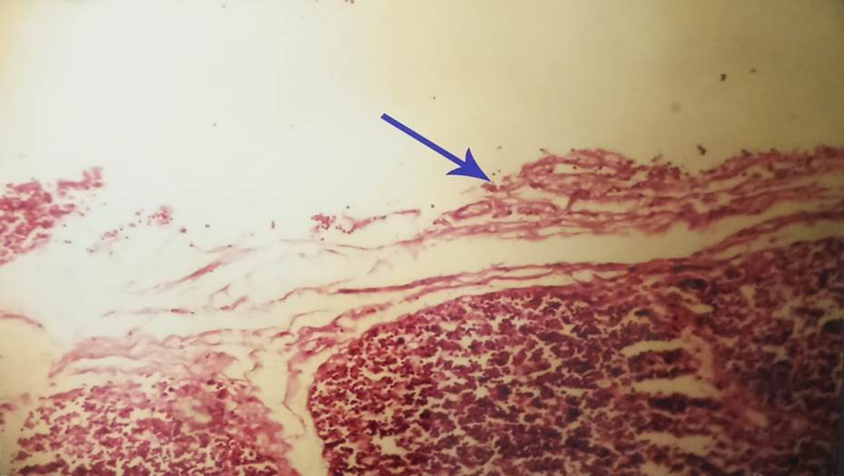
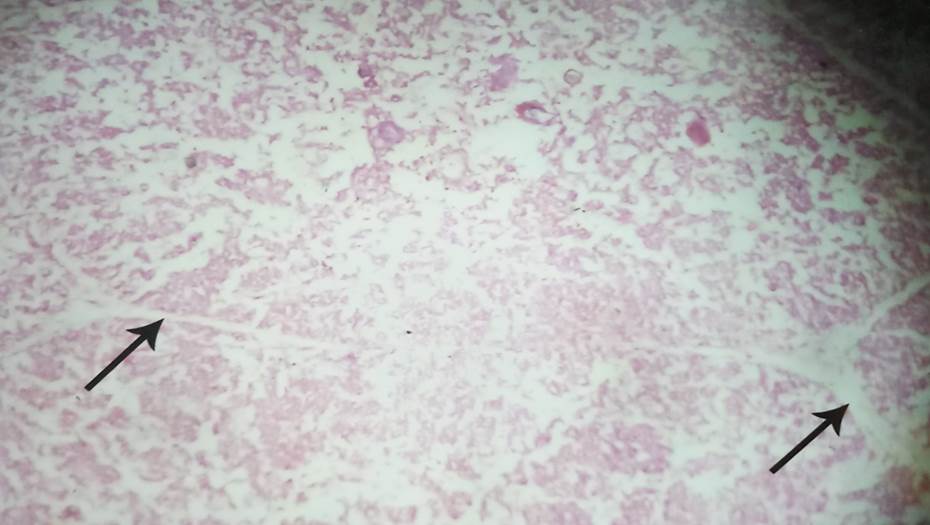
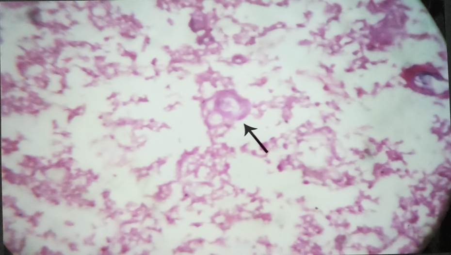
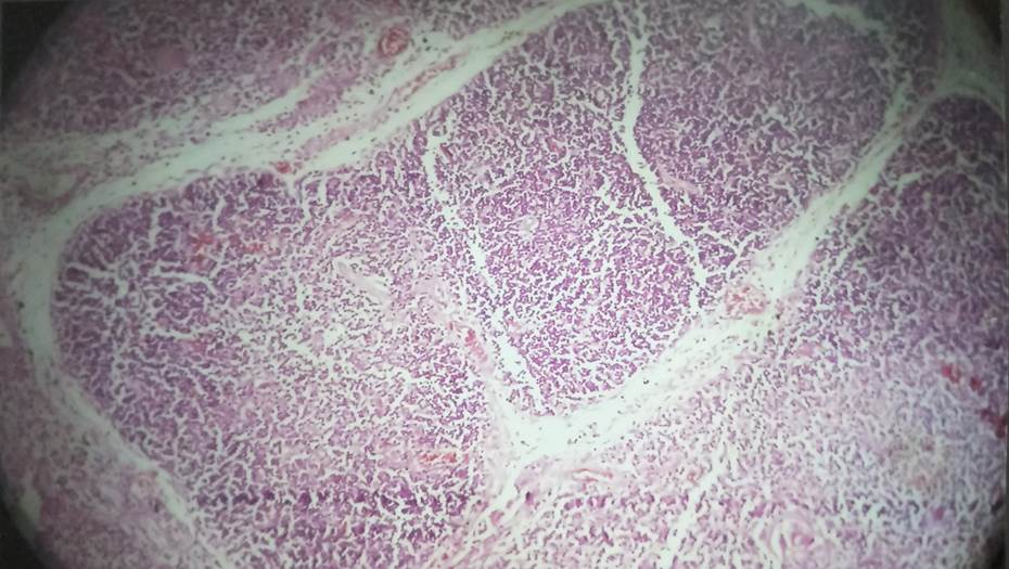
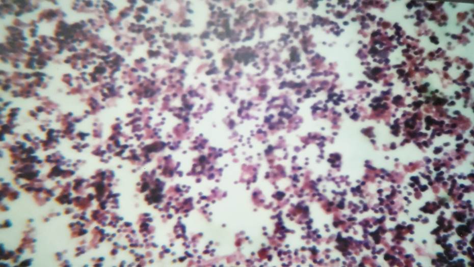
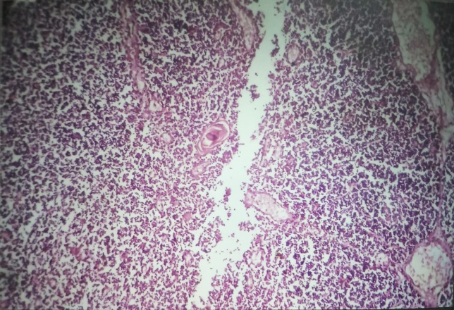
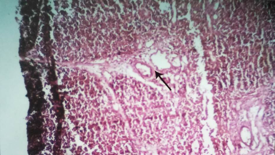
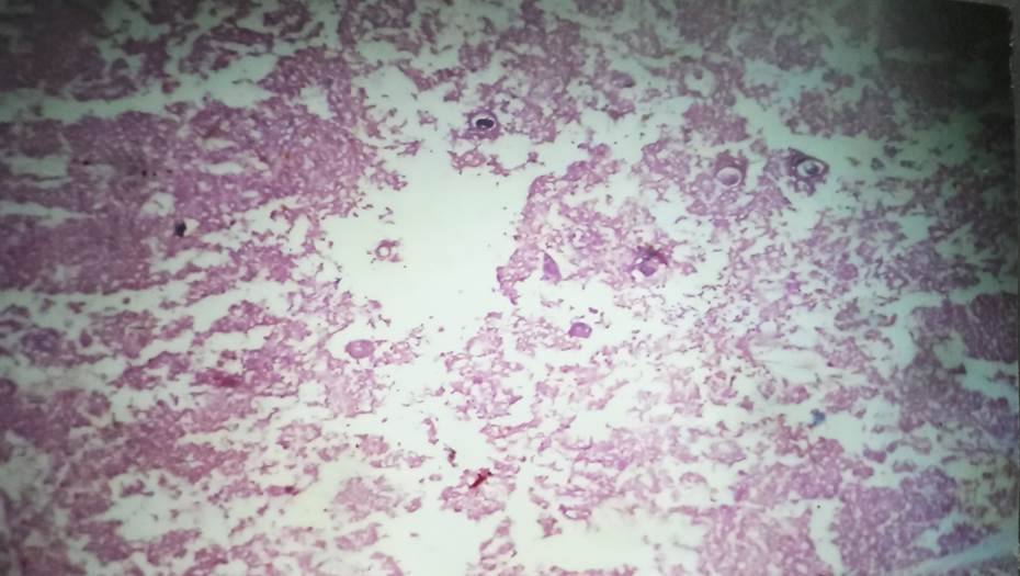
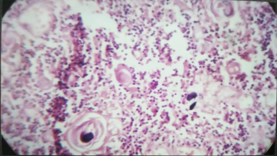
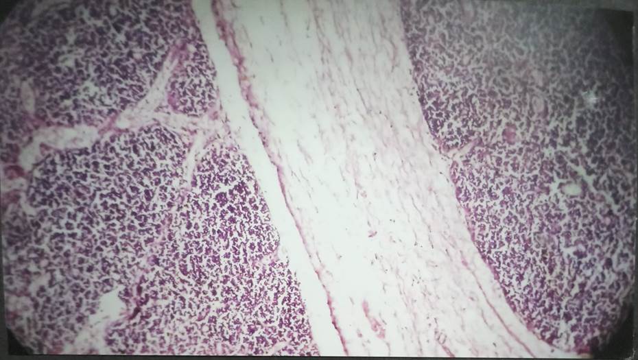
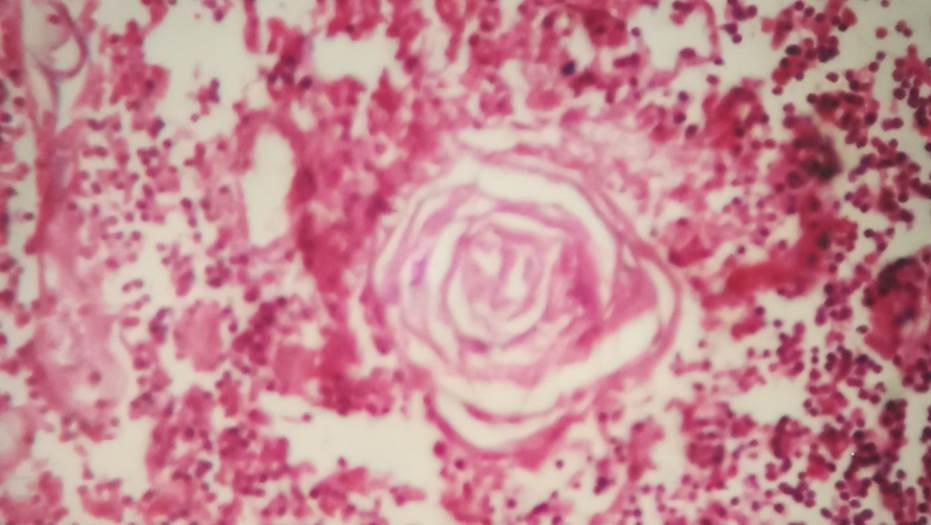
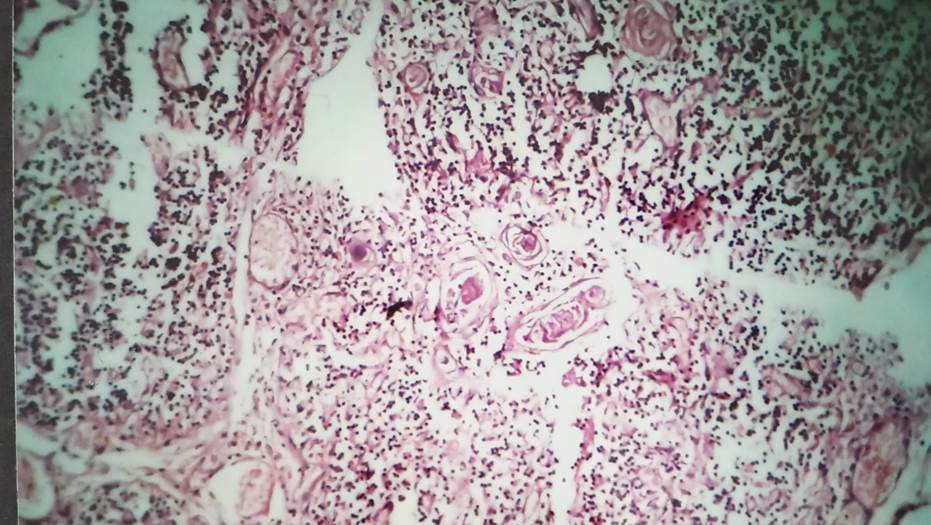
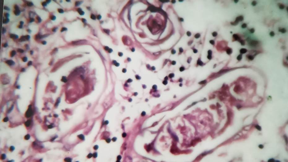
The connective tissue septa had been wider at 18th week and at this degree the lobulation remains endured. Corticomedular differentiation continued to persevere. With many techniques ranging among the lymphocytes, epithelial cells had been abnormal in shape. Few tiny corpuscles of Hassall were evident as concentrated epithelial cells with an imperative eosinophilic mass in the medulla ([Figure 6], [Figure 7], [Figure 8]). Different lobulations changed to visible at 22nd week. The thicker septal connective tissue penetrates deeper into the gland material. Lobules extended in size, with lobulations increasing. At this stage, corticomedular differentiation changed to whole. Numerous and densely packed lymphocytes forming darkly stained cortex were seen at the outer edge of the lobules. Fewer lymphocytes were present at the center and gently stained medulla shaped. Continuous medulla, from one lobule to another ([Figure 9]). Larger blood vessels were seen inside the surrounding connective tissue capsule at 24th week, trabeculae & thymus parenchyma ([Figure 10]), many hassalls corpuscle seen at different stages of development ([Figure 11]). Juvenile type composed of 1 or 2 reticuloepithelial hypertrophic cells. Premature form composed of hypertrophic cell small businesses exhibiting early keratinization techniques, but without a flattened dimension, or a concentration-disposition bent. The reticulo-epithelial cells appear dense at mature level and dispose concentratively of roughly keratin and a mixture of degenerated lymphocytes and macrophages, with or without empty space. Several juvenile type corpuscle of Hassall, some of premature form and mature type have been found ([Figure 11]) at this stage the gland appears to be fully distinguished.
Group-II (25-29 weeks)
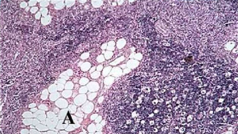
At this degree the connective capsule tissue protecting the gland became very much. At this point, septa (trabeculae) became vast. Connective septa tissue (trabeculae) has also turned thin and fine. At this degree, cortex and medulla have become very gorgeous. Inside the cortex, there was a dense population of lymphocytes, and fewer in the medulla. Simple demarcation between medulla and cortex was evident ([Figure 12], [Figure 13]). Larger blood vessels were evident within the septum of the connective tissue. Smaller blood vessels at corticomedular junction were also visible. The blood vessels identified were of large length than the previous group. Blood vessels had also been found to be more extreme and large. Epithelial cells were anomalous in shape, with many network-forming procedures. Numbers of the corpuscle of Hassall were more than of the previous stage. Many forms of Hassall's corpuscle consisting of young, prema ture, adult, and advanced were seen. Advanced form of Hassall's corpuscle consists of varying degrees of material deposition at its middle or outer edge, while other HC with a distorted form seemed to attempt to fuse with other nearby HC ([Figure 13], [Figure 14]).
Group-III (30-35 weeks)
The gland had an internal architecture visible in adults at some point of this stage, with mature histological picture. Large lobulae have been found. At this degree, the trabecular system became ever cool. At this point the corticomedullary junction was clearly located. At this point, mature, and large blood vessels were evident. Blood vessels with mature vascular frame were elevated in quantities ([Figure 15], [Figure 16], [Figure 17], [Figure 18]). Inside the medulla were present numerous corpuscles of Hassall. Various types had been evident, such as Juvenile, Premature, Mature, and Advanced. At this stage, advanced kind of Hassall's corpuscle was more numerous.
Group-IV (Thymus gland in 15 yrs adolescent)
The gland had an internal architecture seen in adults during this stage, with mature histological image. Lobules were detected to a larger size. At this stage, the trabecular framework became ever more distinct. At this stage corticomedullary junction was clearly observed. This stage saw expanded and larger blood vessels. Blood vessels with mature vascular frame were increased in number ([Figure 19], [Figure 20]). In the medulla were several corpuscles of Hassall’s.

Discussion
Lobulations
Lobulations were present at 12th week in the present study, although lobulations persisted at 12th week level. Distinct lobbing present at the stage of the 18th week. Researchers found evidence that the lobulation was observed at 10th week[12] and that the lobulation was confirmed at 12th week.[13] Therefore, these observations show that lobula formation started at week 9 and distinct lobulae were seen at week 12.[14] These experiments documented that the interlobular septa was noticeably broad at 14-16 weeks; Krishnamurthy observed it at 16 weeks, while the present study was observed at 14-18 weeks.[15]
Cortex and Medulla
In support of this concept, it has been shown that the differentiation between the cortex and the medulla was noticed during the 12th week but now it has not been much distinguished and it has begun to differentiate from the 14th week stage onwards, which has been completed at the 18th week. It has been demonstrated in support of this concept that it became mentioned at the thirteenth week.[13], [16] Observation of using these at 14th week. Between the 12th[17] and 14th weeks[18] it was recorded that cortex and medulla differentiation began at the 9th week and becomes distinct at the 12th to the 14th week.
Blood vessels
Blood vessels were found at 12th week in the present study, because the present study examined fetuses from 12th week onwards, it could not be ascertained when blood vessels begin to appear before 12th week. In second group they became larger, more mature and more numerous. Reported at 11th week of gestation thymus was vascular.[12] Reported that at 9th week there were extrathymic blood vessels associated with connective tissue fibers and mesenchymal cells circling the thymus.[13] Williams et al.[11] listed the 10th week old thymic tissue of the developing erythroblastic cell.[19]
Epithelial cells
The epithelial cells were seen from this week on, as the present review accomplished from 12th week onwards only. Because fetuses were no longer studied within the present week, it could not be determined whether or not there were epithelial cells in the earlier stages. The investigators suggested that the presence of epithelial cells from 8th week[19] was described, and the epithelial component of thymus at 10th week[20] was identifiable.
Hassall’s corpuscles
The corpuscle of the hassall was first observed in the present study at the 12th week of gestation. The Corpuscles of hassall grew in number and size during the 18th to 24th week. The corpuscles of these hassall had variable sizes, ranging from very small to very large whereas early-age fetuses represented the smallest size. Hassall's corpuscles differed considerably in form. Different types such as adolescent, young, adult, advanced observed irrespective of gestational age, with advanced type more notably observed in fetuses over 28 weeks gestational. This polymorphic activity of hassall's corpuscle found in this study correlates with findings that recorded the existence of Hassall's Corpuscles as early as the 8th week of gestation, from the 9th week[21] and at the 11th week, sawant[22] reported the presence of hassall's Corpuscles from the 15th week of gestation. Observations reveal that the Corpuscles of hassall increased in number and size during the 17th to 24th week.[15]
Completion of differentiation
All important structural changes in thymus occur within 18th week of gestation in the present study. Additional evidence for that distinction was completed at week 18,[22] noting that at week 17 thymus appeared to be completely differentiated.[14]
Conclusion
Thymus has a completely unique morphology and histology in distinct age groups. As the CRL will increase there is boom inside the size of the gland. The weight of the thymus gland becomes made as an instantaneous estimate in this study. It was discovered to steadily increase after duration of viability. This becomes justified by means of the fine Pearson correlation and their importance’s become established. The capsule was determined at 12th weeks. The considerable septal formation and deposition of adipocytes within the parenchyma came about with advancement in age of the fetuses thymus. The cortex is densely populated with lymphocytes when as compared with medulla. As age advances the demarcation between the two is step by step lost. The concentric sort of Hassall's corpuscles modified to cystic kind in post viable period. Limitation has been that a small quantity of fetuses was used for this study. Yet studying the thymus gland at special gestational age will essentially give vital insights and will also help us to improve the application of those results. The involution of thymus gland, pondered in its morphology and histology can serve as a baseline to perceive many diseases and may be used as a supplement in screening the growing fetuses. This being a non-invasive approach can also be thoroughly and extensively used to foretell the wellbeing of the unborn fetuses.
Acknowledgment
Authors are thankful to the Principal, Kakatiya Medical College, Telangana, India for his guidance, kind help and constant encouragement at every step during the progress of my work without which successful completion of this work wouldn't have been possible.
Source of Funding
None.
Conflict of Interest
The authors declare that they have no conflicts of interest.
References
- Bellamy D. The Thymus in Relation to Problems of Cellular Growth and Ageing. Gerontol. 1973;19(3):162-84. [Google Scholar]
- Gui J, Mustachio LM, Su DM, Craig RW. Thymus size and age-related thymic involution: early programming, sexual dimorphism, progenitors and stroma. Aging Dis. 2012;3(3):280-90. [Google Scholar]
- Aw D, Silva AB, Maddick M, Zglinicki Tv, Palmer DB. Architectural changes in the thymus of aging mice. Aging Cell. 2008;7(2):158-67. [Google Scholar]
- Lynch HE, Goldberg GL, Chidgey A, Brink MRVd, Boyd R, Sempowski GD. Thymic involution and immune reconstitution. Trends Immunol. 2009;30(7):366-73. [Google Scholar]
- Gewolb IH, Lebowitz RL, Taeusch HW. Thymus size and its relationship to the respiratorydistress syndrome. J Pediatr. 1979;95(1):108-11. [Google Scholar]
- Gui J, Mustachio LM, Su DM, Craig RW. Thymus size and age-related thymic involution: early programming, sexual dimorphism, progenitors and stroma. Aging Dis. 2012;3(3). [Google Scholar]
- Baron RL, Lee JK, Sagel SS, Peterson RR. Computed tomography of the normal thymus.. Radiol. 1982;142(1):121-5. [Google Scholar]
- Francis IR, Glazer GM, Bookstein FL, Gross BH. The thymus: reexamination of age-related changes in size and shape. Am J Roentgenol. 1985;145(2):249-54. [Google Scholar]
- Baron RL, Lee JK, Sagel SS, Levitt RG. Computed tomography of the abnormal thymus.. Radiol. 1982;142(1):127-34. [Google Scholar]
- Moore AV, Korobkin M, Olanow W, Heaston DK, Ram PC, Dunnick NR. Age-related changes in the thymus gland: CT-pathologic correlation. Am J Roentgenol. 1983;141(2):241-6. [Google Scholar]
- Hasselbalch H, Jeppesen DL, Ll AKE, Engelmann MD, Nielsen MB. Thymus size evaluated by sonography: a longitudinal study on infants during the first year of life. Acta Radiol. 1997;38(2):222-7. [Google Scholar]
- Ghali WM, Abdel-Rahman S, Nagib M, Mahran ZY. Intrinsic innervation and vasculature of pre- and post-natal human thymus. Cells Tissues Organs. 1980;108(1):115-23. [Google Scholar]
- Haar JL. Light and electron microscopy of the human fetal thymus. Anat Rec. 1974;179(4):463-75. [Google Scholar]
- Ajita RK, Singh TN, Singh YI, Singh LC. An insight into the structure of the thymus in human foetus-a histological approach. J Anat Soc India. 2006;55(1):45-9. [Google Scholar]
- Krishnamurthy JV, Devi VS, Reddy JV. Developmental histology of human foetal thymuses at different gestational ages. J Evol Med Dent Sci. 2015;4(40):6944-54. [Google Scholar]
- Varga I, Pospisilova V, Jablonska-Mestanova V, Galfiova P, Polak S. The thymus: picture review of human thymus prenatal development. Bratislavske Lekarske Listy. 2011;112(7):368-76. [Google Scholar]
- Lobach DF, Haynes BF. Ontogeny of the human thymus during fetal development. J Clin Immunol. 1987;7(2):81-97. [Google Scholar]
- Gaudecker B. Functional histology of the human thymus. Anat Embryol. 1991;183(1):1-5. [Google Scholar]
- Williams P, Dyson M, Dussak JE, Collins P, ed 3. . Gray's Anatomy. Skeletal system. 1995. [Google Scholar]
- Hayward AR. Myoid cells in the human foetal thymus. J Pathol. 1972;106(1):45-8. [Google Scholar]
- Gilhus NE, Matre R, Tönder O. Hassall's corpuscles in the thymus of fetuses, infants and children: immunological and histochemical aspects. Thymus. 1985;7(2):123-35. [Google Scholar]
- Sawant SP. Development of thymus. J Anat Soc India. 2003;52(1). [Google Scholar]
- Abstract
- Introduction
- Materials and Methods
- Results
- Measurement
- Weight
- In lobule
- Histology
- Group-I (18-22 weeks)
- Group-II (25-29 weeks)
- Group-III (30-35 weeks)
- Group-IV (Thymus gland in 15 yrs adolescent)
- Discussion
- Lobulations
- Cortex and Medulla
- Blood vessels
- Epithelial cells
- Hassall’s corpuscles
- Completion of differentiation
- Conclusion
- Acknowledgment
- Source of Funding
- Conflict of Interest
- References
Article Metrics
- Visibility 1.2k Views
- Downloads 253 Views
- DOI 10.18231/j.ijcap.2020.045
-
CrossMark

- Citation
- Received Date November 30, -0001
- Accepted Date November 30, -0001
- Publication Date June 27, 2020