Indian Journal of Clinical Anatomy and Physiology
Indian Journal of Clinical Anatomy and Physiology (IJCAP) is an open access, peer-reviewed medical quarterly journal, published since 2014 under the auspices of the Innovative Education and Scientific Research Foundation (IESRF), which aims to uplift researchers, scholars, academicians, and professionals in all academic and scientific disciplines. IESRF is dedicated to the transfer of technology and research by publishing scientific journals, research content, providing professional memberships, and conducting conferences, seminars, and award programs....
A study on morphometric dimensions of tibial condyles in Indian population
Abstract
Background and Aim: Knee joint is the largest and complex joint in the body which helps in weight bearing and locomotion. It is subjected to degenerative changes like osteoarthritis, Knee injuries and trauma. Present study is proposed to access measurements of upper end of tibia. These morphometric parameters can be useful to guide the treatment in procedures of unicompartmental knee arthroplasty and total knee arthroplasty.
Material and Methods: Present study was conducted in Index Medical College, Department of anatomy, Indore, Madhya Pradesh. Different parameters of 62 human dried tibiae [31 right and 31 left] measured are anteroposterior [AP] and tranverse diameter [TD] of medial condyle, lateral condyle and total tibial condyle.
Results: In present study, mean values of anteroposterior and transverse diameters of medial condyle, lateral condyle and total tibial condyle were found to be slightly greater on left side than that of right side. Furthermore AP diameter of medial condyle were found to be greater than that of lateral condyle on both sides and TD of medial found to be lesser than that of lateral on both sides.
Conclusion: Results of this study will be helpful for orthopedicians to design appropriate size of knee prosthesis in knee arthroplasty also useful for forensic experts, anthropologist and anatomist.
Introduction
Knee joint is the largest joint in the human body. It is a compound synovial joint, performs a significant role in adjusting the centre of body mass and posture which requires a great range of movements in three dimensions together with the capability to bear high forces.[1]
Knee joint is a modified hinge joint, composing of tibofemoral and patellofemoral components. The main articular surface of femur and tibia are its medial and lateral condyles in its lower and upper end respectively. The pair of tibial condyles are separated by the intercondylar eminence. The articular surface of lateral condyle is circular while medial condyle is oval. Through the tibiofemoral articulation, the upper end of tibia plays a crucial role in conducting the body weight from femur above to talus below, thus being unique in performing day to day activities and sports.[2] Therefore knee joint is commonly affected by osteoarthritis which is the most common pathological disorder of knee. Osteoarthritis is usually treated by Total Knee Arthoplasty [TKA] or Unicompartmental Knee Arthoplasty[3] [UKA]. In TKA precise soft tissue balancing is required along with resection of bone thickness equal to the thickness of prosthetic component implanted which helps in equal spacing for flexion & extension and permits joint stability throughout the range of motion. The success of TKA depends on the prosthetic selection, accurate sizing and proper placement. In order to sustain the flexion & extension spacing, Anteroposterior measurements [AP] of prosthetics is essential. The mediolateral measurements decide essential coverage of resected bone surface and wound closure.[4] In UKA, damaged knee parts are replaced only in a single compartment.UKA is designed to cause less trauma and damage by replacing only less bone and maintaining most of the individuals own bone.[5] At present UKA is a satisfying treatment for unicompartmental knee arthritis. The prosthetics are made with the measurements available from western population studies. Hardly few Indian studies are available on the anthropometric measurements of upper end of tibia. Hence this study aims to measure the dimensions of upper end of tibia which can be a useful guide for designing knee prostheses for Indians.
Researchers also use Maximum Tibial Length [MTL] for estimating human stature. Estimating human stature with MTL is of great use in forensic medicine and physical anthropology.[6]
Materials and Methods
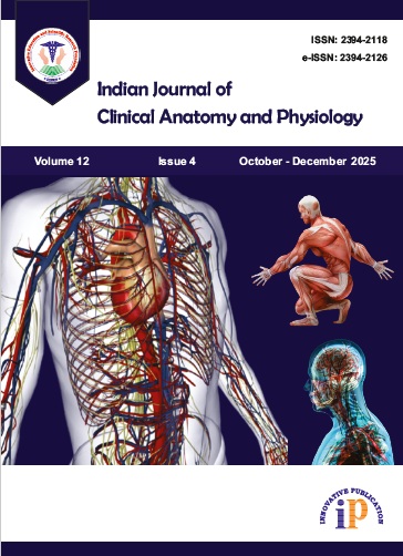
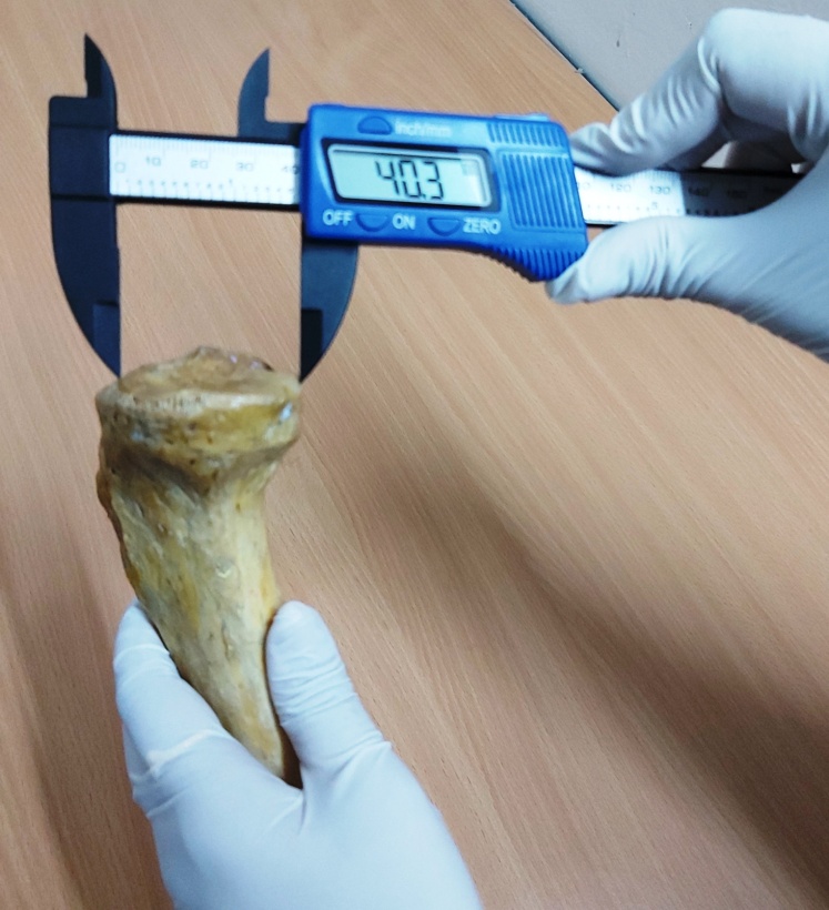
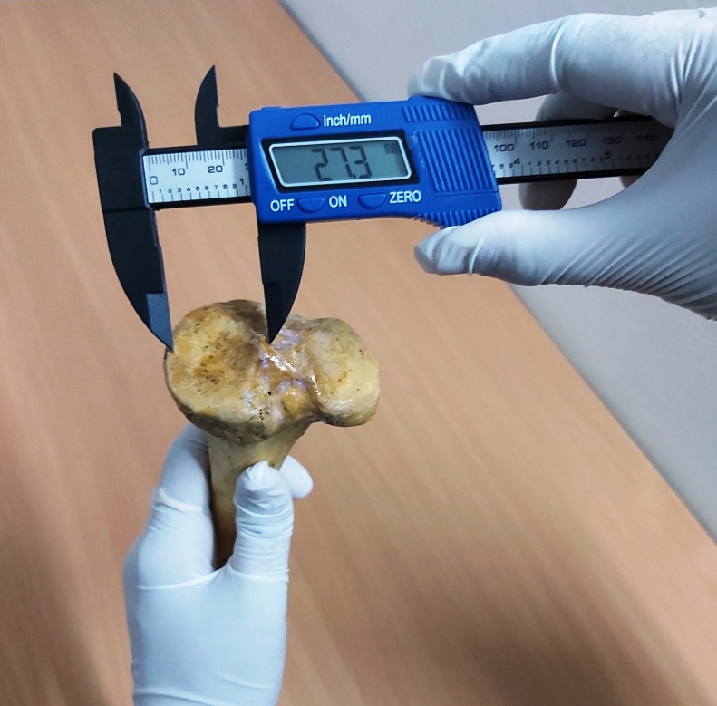
The present study was conducted in Department of Anatomy, Index Medical College and Research Centre, Indore, Madhya Pradesh. 62 dried human tibiae bones [31right & 31 left] of unknown age and sex were analysed. Samples were obtained from Index medical college and Sri Aurobindo Institute of Medical Sciences, Indore, Madhya Pradesh. Damaged, fractured and unossified bones were excluded from the study. Using standard digital vernier caliper dimension values were measured in millimeter (mm). The dimensions to be measured for conducting present study are Medical Condyle Anteroposterior diameter [MCAP], Lateral condyle Anteroposterior diameter [LCAP], Medical Condyle Transverse diameter [MCTD], Lateral Condyle Transverse diameter [LCTD], Total tibial condyle Anteroposterior [TTC-AP] and total tibial condyle Transverse diameter[TTC-TD]. MCAP was measured from the most anterior point to most posterior point as a single line. MCTD is measured from the medial tubercle to medial margin of medial condyle as a straight line perpendicular to AP diameter. LCAP was measured from the most anterior point to most posterior point as a single line. LCTD is measured from the lateral tubercle to lateral margin of lateral condyle as a straight line perpendicular to AP diameter. AP diameter of TTC is measured as straight line from the middle of medial and lateral tubercle of intercondylar eminence from the most anterior to posterior point. Transverse diameter of TTC is measured as a straight line from the medial margin of medial condyle to lateral margin of lateral condyle. The measurements were performed by same person to avoid inter observer errors. Data obtained were tabulated and statistically analyzed using SPSS software (version 12.0). Values between right and left sides tibiae were statistically analyzed using unpaired t- test, whereas values between the medial and lateral condyle statistical analyzed by using paired t test. p value less than 0.05 is considered statistically significant.


Results
The morphometric study is conducted on 62 human dried tibiae, all measured values of different parameters are tabulated, analyzed and subjected to statistical analysis by using SPSS software (version 12.0). The results are as follows:
| Side of tibia | Medial condyle | Lateral condyle | Total tibial condyle | |||
| MCAP | MCTD | LCAP | LCTD | TTC-AP | TTC-TD | |
| Right | 39.38 ± 4.08 | 27.64 ± 2.64 | 34.81 ± 3.76 | 28.52 ± 3.28 | 43.58 ± 3.89 | 63.89 ± 7.29 |
| Left | 40.08 ± 3.44 | 29.11 ± 3.14 | 35.42 ± 3.33 | 29.47 ± 3.01 | 45.03 ± 3.71 | 66.22 ± 4.38 |
| Parameter | Range Right(Mean ±SD) | Range left (Mean ±SD) |
| Medial condyle AP length | 39.38 ± 4.08 | 40.08 ± 3.44 |
| Lateral condyle AP length | 34. 81 ± 3.76 | 35.41 ± 3.33 |
| Medial condyle TD length | 27.64 ± 2.64 | 29.11 ± 3.14 |
| Lateral condyle TD length | 28.52 ± 3.28 | 29.54 ± 3.06 |
| Total tibial condyle AP Length | 43.58 ± 3.89 | 45.12 ± 3.66 |
| Total tibial condyle TD length | 63.89 ± 7.29 | 66.22 ± 4.38 |
[Table 1], [Table 2], the Total Tibial Condyle (TTC) area is calculated as: TTC = AP × TD,[7] So TTC area of right side is 2784.33 mm and TTC area of left side is 3120.28 mm.
As shown in the [Table 1], the mean values of TTC- AP are 43.58 ± 3.89 mm on right side while on the left it was 45.03 ± 3.71 mm and the mean values of TTC -TD was 63.89 ± 7.29 mm on right side while on the left side it was 66.22 ± 4.38 mm; so the difference is considered as statistically not significant [p value = 0.13]. The mean values of medial condylar AP length on right side comes to be 39.38 ± 4.08 mm and on left side it was 40.08 ± 3.44 mm. The mean values of medial condylar TD length on right side comes to be 27.64 ± 2.64 mm and on left side it was 29.11 ± 3.14 mm which is considered to be not statistically significant.
The mean values of lateral condylar AP length on right side comes to be 34.81 ± 3.76 mm and on left side it was 35.42 ± 3.33 mm. The mean values of lateral condylar TD length on right side comes to be 28.52 ± 3.28 mm and on left side it was 29.47 ± 3.01 mm which is considered to be not statistically significant.
According to [Table 2], it was observed that mean value of MCAP was 39.38 ± 4.08 mm and LCAP was 34.81 ± 3.76 mm on right side. The p value is less than 0.0001, this difference is considered to be extremely statistically significant. Similarly the mean value of MCTD was 27.64 ± 2.64 while LCTD was 28.52 ± 3.28 mm [p = 0.0229] this difference is considered to be statistically significant.
It was observed that mean value of MCAP was 40.08 ± 3.44 mm and LCAP was 35.41 ± 3.33 mm on left side. The p value is less than 0.0001, this difference is considered to be extremely statistically significant. But it was seen that the mean value of MCTD on left side was 29.11 ± 3.14 mm while LCTD was 29.54 ± 3.06 mm.
As per table no. 2, mean values of TTC TD was 63.89 mm which is greater than TTC- AP= 43.58 mm. that is on right side, while mean values of TTC- TD was 66.22 ± 4.38 mm which is greater than TTC AP = 45.12 ± 3.66 mm. that is on left side, P value is less than 0.0001 and this difference is considered to be statistically significant.
Discussion
From past, studies done by various authors on different populations across India and abroad it was recorded that Indians need smaller size knee prosthesis as compared to Caucasian and western population. Even though in past, many studies conducted by CT and MRI, has been reported inaccurate. So direct measurements taken from dry bones have significant advantage in designing knee prosthesis.
The study from P Chaitanya Shree et al[7] which was based on Tamil Nadu population showed that the medial condylar AP length was 37.91mm was greater than the lateral one which was 36.89mm. From another study done by Ankur Zalawadia[8] it was seen that the Medial condylar AP length 44.27 ± 1.93 mm greater than that of lateral condylar AP 38.26 ± 2.43 mm. This study was based on Gujarat population. The present study was highly correlated with the aforementioned studies. The mean values of medial condylar AP length were 39.38 ± 4.08mm was greater than the lateral one which was 34.81 ± 3.76 mm.
The study done by Swati Gandhi[9] who had measured 100 adult human dry tibiae, 50 male and 50 female, in Indian population. AP measurements of intercondylar area were 47.19 ± 2.93 mm on right side and 49.11 ± 3.97 mm on left. Our measurements correlated with this study as TTC AP was 43.58 ± 3.89 mm on right and on left it was 45.03 ± 3.71 mm. our study also compared with Chandni Gupta et al,[2] in Karnataka population, AP length of intercondylar region was found as 46.6 mm on right side and 44.9 mm on left side and Transverse diameter of tibial condyle were 67.7 ± 0.31 mm on right side and left side 68.8 ± 0.65 mm, our study almost correlated with aforesaid study, as TTC TD on right side 63.89 ± 7.29 mm and on left side 66.22 ± 4.38 mm.
Bukkumbuddhi Virupakshamurthy Murlimanju[10] et al. studied 73 cadaveric tibiae on South Indian Population. Breadth of medial tibial plateau was 26.7 mm and of lateral tibial plateau 26.1 mm. In our study, MCTD were 27.64mm and LCTD 28.52 mm, which was highly correlated.
Ankit Srivastava et al.[11] measured 150 dry tibiae. The mean area of TTC was 2988.73 on right and on left 2951.44 mm. In present study, Total Tibial Condylar areas were 2784.33mm on right side and on the left side were 3120.28mm. Our values were found to be greater than above mentioned study.
Conclusion
Tibial condylar shape gains an importance because it provides attachment to medial and lateral menisci. As per study reports, menisci occupies 55–66.7% of the corresponding tibial condyles.[11] Up to 14th week of intrauterine life tibial condyles and menisci are of same size on both sides. During later stage of development the size of menisci increases. However the medial condylar area increases more rapidly because of the smaller coverage area of the medial menisci.[12] So the measured parameteric values could be helpful for meniscal transplantation.[13] Osteoarthrits of knee joint is very common degenerative disease in adults. It is mainly due to more mechanical stress and genetic factors.[14]
This study will be highly beneficial for orthopeadic surgeons for designing appropriate sizes of knee prosthesis in unicompartmental and also in total knee arthroplasty. Study is also helpful forensic experts, anthropologist and anatomist.
Source of Funding
None.
Conflict of Interest
None.
References
- SS. . Gray's Anatomy: The Anatomical Basis of Clinical Practice. 2005. [Google Scholar]
- Gupta C, Kumar J, Kalthur SG, D′souza AS. A morphometric study of the proximal end of the tibia in South Indian population with its clinical implications. Saudi J Sports Med. 2015;15(2). [Google Scholar]
- Ivan AS. Morphometric Study of Proximal End of Tibia. . 2014. [Google Scholar]
- Vaidya SV, Ranawat CS, Aroojis A, Laud NS. Anthropometric measurements to design total knee prostheses for the Indian population. J Arthroplast. 2000;15(1):79-85. [Google Scholar]
- Borus T, Thornhill T. Unicompartmental Knee Arthroplasty. J Am Acad Orthop Surg. 2008;16(1):9-18. [Google Scholar]
- Attada K, Veera P, Ravindranadh G, Kumari U, Deena K. Anthropometric Measurement of Maximum Tibia Length in South Indian Population. Anat Sci J. 2017;14(2):47-54. [Google Scholar]
- Shree PC, Babu KY, Mohanraj KG. Morphological and morphometric study of tibial condylar area and its clinical significance. Drug Invention Today. 2018;10. [Google Scholar]
- . Morphometric study of upper end. Int J Anat Res. 2018;6(1):4836-9. [Google Scholar]
- Gandhi S, Singla RK, Kullar JS, Suri RK, Mehta V. Morphometric analysis of upper end of tibia. J Clin Diagnostic Res. 2014;8(8). [Google Scholar]
- Murlimanju BV, Purushothama C, Srivastava A, Kumar CG, Krishnamurthy A, Blossom V. Anatomical morphometry of the tibial plateau in South Indian population. Italian J Anat Embryol. 2016;121(3):258-64. [Google Scholar]
- Srivastava A, Yadav A, Thomas RJ, Gupta N. Morphometric study of tibial condylar area in the North Indian population. J Med Sci Clin Res. 2014;2:515-24. [Google Scholar]
- Fukazawa I, Hatta T, Uchio Y, Otani H. Development of the meniscus of the knee joint in human fetuses. Congenital Anomalies. 2009;49(1):27-32. [Google Scholar]
- Malegaonkar SS, Malegaonkar DB, Naik S, Shireen S. A Morphometric Study of the Proximal End of Tibia in North East Karnataka Population with Its Clinical Implication. Indian J Anat. 2017;6(1):81-5. [Google Scholar]
- Nayak G, Panda SK, Chinara PK. Morphometric analysis of tibial plateau. Int J Res Med Sci. 2019;7(4). [Google Scholar]
Article Metrics
- Visibility 716 Views
- Downloads 129 Views
- DOI 10.18231/j.ijcap.2020.056
-
CrossMark

- Citation
- Received Date November 30, -0001
- Accepted Date November 30, -0001
- Publication Date October 17, 2020